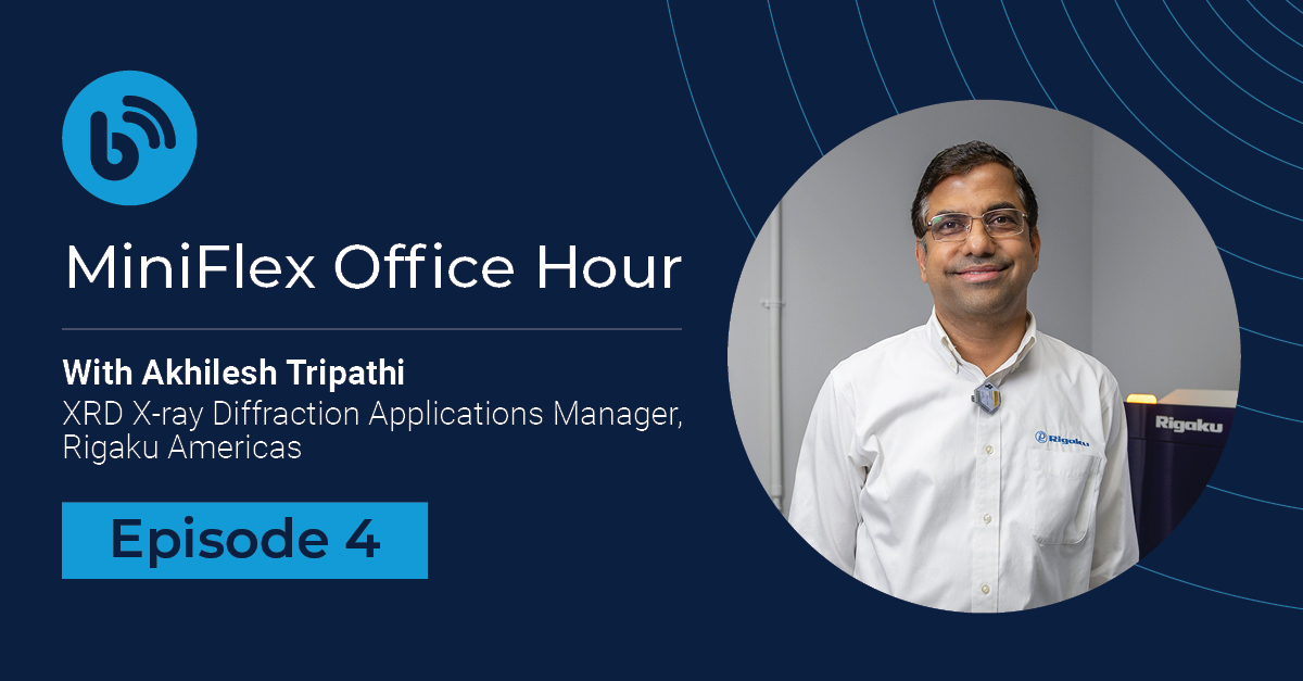
MiniFlex Office Hour Episode # 4 Recap
Jun 5, 2025

Recap provided by Aya Takase
Thank you to everyone who joined us for Episode 4 of MiniFlex Office Hour! As always, our XRD expert, Akhilesh Tripath, shared valuable insights and answered practical questions from MiniFlex users around the world. Below is a quick recap of the key topics we covered.
You can watch the full recording here. If you’re new to MiniFlex, it’s a benchtop X-ray diffractometer that researchers have trusted since 1973.
Episode recap
-
Liquid sample analysis using XRD is possible, particularly for mixtures that aren't fully dissolved (such as slurries) and viscous liquids, such as petroleum products.
To handle these, a specialized liquid sample holder is used. It includes an O-ring and a small hole at the back. First, seal the holder using a polymer or Mylar film. Then, insert the O-ring using the designated tool, pour in the liquid sample (typically with a syringe), and secure the back with tape. You can trim any extra film if needed.
Once prepared, the holder fits into the ASC-8 sample changer.
(Watch Akhilesh demonstrate the liquid sample holder in the episode recording.)
-
Chiller water maintenance depends on chiller type and lab conditions. For external chillers, check the water every 3 to 6 months, or more frequently in hot environments. If the water level is below the marked line, add water. If the water is murky, replace the water. Remember to use distilled (not deionized) water.
Internal chillers on newer MiniFlex units are more robust, and Rigaku provides additives to help prevent issues such as algae buildup, so you don't need to check them as frequently.
Please note that if the water flow drops below the threshold, the protection circuit kicks in and the X-rays will automatically shut off. So, you don't need to worry about damaging the X-ray tube, although you should check the water condition before the safety circuit is triggered.
-
I recommend checking the data integrity monthly or bi-weekly using a standard like NIST SP 640G. Perform unit cell or Rietveld refinements and compare results to official values, and they are accurate to two decimal places. If the value is off, you should rerun the instrument alignment and calibration.
-
Older versions don’t reduce result accuracy. Updates mostly bring bug fixes and new features, such as advanced analysis methods. It's good to check with your sales representative if upgrades offer value for your work.
-
Errors arise in three key areas: sample preparation, data collection, and analysis.
Sample preparation:
Ensure the crystallite or grain size is below 40 microns using sieves or thorough grinding.
Make sure the sample is packed properly. Poor packing causes displacement errors. Samples packed too high shift peaks high in 2θ, and too low shift them low. Pack flush with the holder’s brim.
Data collection:
I recommend a quick test scan to figure out the optimum scanning conditions, such as the step size, speed, and range.
If you don't have time to run a test scan, 0.02° is a good default. If you have time, get the FWHM from the test scan and divide it by 5–7 to determine a suitable step size.
Set the scan speed to run for about 10 minutes for most samples if you have a high-speed detector. Adjust it to achieve the required data quality for the analysis.
For the range, Inorganics typically need a 3–90° 2θ range; organics can use 3–50°.
Analysis:
Use a good database and hone your interpretation skills. It is important to use your common sense and knowledge about the sample to rule out potential incorrect phase identification. Identifying multiple phases accurately or doing quantitative Rietveld analysis requires more expertise than basic phase identification.
-
Switching to a cobalt tube helps with fluorescence issues in iron-rich samples. When using cobalt, the XRD software automatically adjusts matching parameters for cobalt radiation, so you don't need to worry about manually adjusting them.
The only manual step is setting the tube type to "Cobalt 2" in the instrument settings. That’s it. The data collection software handles everything else.
I want to add a comment about the systems with the standard copper tube. If you’re using a copper tube but have moderate iron content (e.g., 50–70%), you can take advantage of the newer detectors' X-ray fluorescence reduction mode. These detectors can reduce the background from iron fluorescent X-rays by narrowing their energy window. They often make a cobalt upgrade unnecessary unless you're analyzing iron-heavy samples regularly.
-
It's strongly recommended to upgrade to the Comprehensive software package. It supports instrument correction, which is critical for consistent and accurate crystallite size estimates.
Previously, lanthanum hexaborate (LaB₆) was the go-to standard, but now Rigaku supports corundum and silicon as alternatives. You collect data on the standard using the same slit setup as your samples. The software then applies corrections based on this to subtract the peak broadening caused by the instrument resolution and calculate the crystallite size purely based on the sample-originated peak width.
-
It’s the powder sample that satisfies Bragg’s law, and the instrument helps it happen and capture a diffraction pattern.
It is common for people to picture a single crystal and wonder how it can satisfy the diffraction condition for so many peaks. However, powder XRD samples are powders and consist of millions of randomly oriented crystallites. When X-rays hit them, some are always positioned just right to meet the Bragg condition (2d sinθ = nλ) for a certain d-spacing, diffraction angle θ, and the X-ray wavelength λ, for n=1, 2, ... As the goniometer scans through angles, different crystallites diffract, generating peaks.
-
In θ-2θ geometry, the incident beam angle (θ) and the detector angle (2θ) maintain a strict 1:2 relationship.
Mechanically, both the detector and the sample rotate while the X-ray source stays still. This method is built to run a scan along the focusing circle of the Bragg-Brentano geometry, making precise diffraction measurements possible while maintaining both high angular resolution and high X-ray intensity.
-
Yes! Both PDXL and SmartLab Studio II (SLS II) support importing XRF data to assist in phase identification.
Under the "element" tab in the search-match function, there's a "Load XRF data" option. You can load XRF data as a CSV easily if it is from a Rigaku instrument. Third-party data needs to follow the same format.
This doesn’t quantify the elements but highlights them in the software as ones that can be included in the sample, ruling out phases that won't make sense. It is a qualitative aid to support more accurate phase matching.
Join us next time
We’ll be back every month with more questions, tips, and insights from Akhilesh. If you use MiniFlex or are curious about benchtop XRD, we hope you’ll join us live next time!
The live event information is posted on our LinkedIn event page.
Got questions in the meantime? Drop your question in the comment section of the most recent episode. Akhilesh will answer them, or we might answer it live during the next office hour.


Subscribe to the Bridge newsletter
Stay up to date with materials analysis news and upcoming conferences, webinars and podcasts, as well as learning new analytical techniques and applications.

Contact Us
Whether you're interested in getting a quote, want a demo, need technical support, or simply have a question, we're here to help.
