Life Science Research
From protein crystallography to molecular imaging
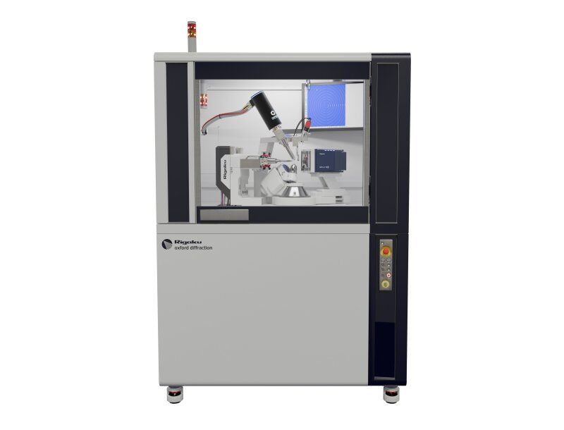
X-ray and electron diffractometers are instrumental in solving protein and small molecule structures for drug discovery and various life science research. Towards the pre-clinical stage, molecular imaging is used to study the organs of small animals. In various natural science studies, X-ray computed tomography (CT) is used to image plants, insects, and soft tissues.
Application notes
Explore the example analyses to see which analytical technique is right for you
-
XCT2201 - Submicron CT for the Life Sciences: Animal and Tissue Research Applications
-
XCT2103 - Submicron CT for Plant Research Applications
-
RACCT9046 - Wild Bird Forensic
-
RACCT9024 - Tomato Plant Root System Analysis by X-ray CT
-
RACCT9023 - Maize Kernel Analysis by X-ray CT
-
RACCT9022 - Coral Surface Area and Porosity Analysis by X-ray CT
-
RACCT9021 - Cicada Analysis by X-ray CT
-
PX031 - Method: MicroED/3D ED on Proteins Using the XtaLAB Synergy-ED
-
PX030 - Using CrysAlisᴾʳᵒ to Tackle Macromolecular Non-merohedral Twins
-
PX029 - Intoducing the XtaLAB Synergy Flow
-
PX028 - Resolving Large Unit Cells by using the Correct Detector, Distance, and Beam Divergence
-
PX027 - Synergy Of PX and SMX on One Diffractometer with the HyPix-Arc 150°
-
PX026 - Sulfur SAD Phasing of Thaumatin in 10 Minutes with the HyPix-Arc 150° and XtaLAB Synergy-DW
-
PX024 - Lysozyme S-SAD Phasing with Two Minutes of Data Collection on the XtaLAB SynergyCustom
-
PX023 - ClpS SAD phasing with 30 Minutes of Data Collection on the XtaLAB Synergy-S
-
PX022 - Thaumatin S-SAD Phasing with 2 hours of Data Collection on the XtaLAB Synergy-S
-
PX021 - I-SAD Phasing of the HIV Integrase Core Domain using Data Collected on the XtaLAB Synergy-S
-
PX020 - In situ Data Collection and Structure Solution using the XtalCheck-S System
-
PX019 - Using the EIGER 4M for Single Crystal X-ray Diffraction on Catalase
-
PX018 - CrysAlisᴾʳᵒ: An All-in-one Software Package for Single Crystal X-ray Diffraction
-
PX015 - Data Collection and Improved Spot Resolution with the HyPix-6000HE HPC Detector
-
B-XRI1017 - Analysis of volume fraction and thickness of bran layer using a high-resolution 3D X-ray microscope
Recommended products
Not seeing what you are looking for? Try Product Finder.
XtaLAB Synergy-S
Our most popular diffractometer for Chemical Crystallography and Mineralogy, configured with either single or dual microfocus sealed tube X-ray sources and an extremely low noise direct X-ray detection detector.
Learn moreXtaLAB Synergy-R
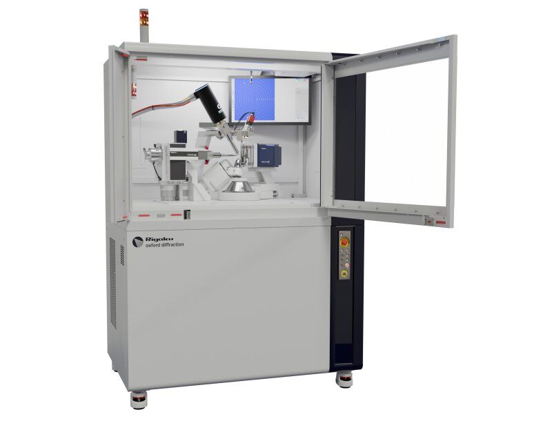
Our most popular diffractometer for Protein Crystallography, configured with a high-flux rotating anode X-ray source and an extremely low noise direct X-ray detection detector.
Learn moreXtaLAB Synergy-ED
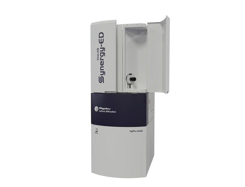
A new and fully integrated electron diffractometer for measuring submicron crystals, utilizing a seamless workflow from data collection to structure determination of crystal structures.
Learn moreXtaLAB mini II
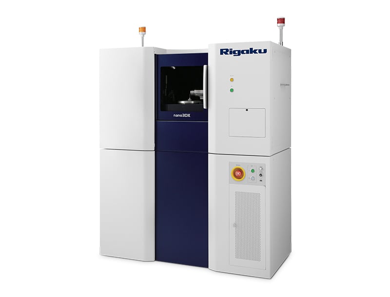
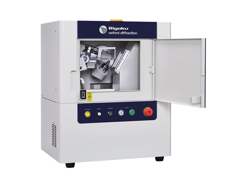
A benchtop single crystal X-ray diffractometer ideal for chemical crystallography and teaching.
Learn moreMILabs - a Rigaku Company Molecular imaging
MILabs is one of the global leaders in molecular imaging and offers high-performance pre-clinical multi-modal imaging systems that can integrate micro-PET, micro-SPECT, micro-CT, and Optical Tomography systems.
Contact Us
Whether you're interested in getting a quote, want a demo, need technical support, or simply have a question, we're here to help.

Subscribe to a Rigaku Newsletter
Stay up to date with the latest Rigaku news!