
Advanced and New Materials
Every aspect of modern life benefits from coating or thin film technology. Whether analyzing a barrier layer film in an integrated circuit chip or a conversion coating on an aluminum beverage can, X-ray analytical techniques are integral to R&D, production process control and quality assurance of such materials. Composition and structure of coatings, thin films, multilayers, materials, solar cells, batteries, semiconductors, nanomaterials and many other materials can be analyzed by X-ray fluorescence (XRF) to determine the thickness and elemental composition in metrology. XRT (X-ray topography) can be employed to investigate crystalline defects and X-ray Reflectometry (XRR) to measure layer thicknesses, roughness and interlayer diffusion. Emerging as a leading enabler for such thin film and nanotechnology research, X-ray diffraction (XRD) and associated diffuse scattering methods like SAXS examine the nature of the molecular structure of films, coatings and layers or particles in biology, chemistry, physics, and engineering. Rigaku’s advanced technology and experience provide a variety of non-destructive and state-of-the-art analytical solutions and service.
Application notes
Explore the example analyses to see which analytical technique is right for you.
-
XRD2001 - Orientation and Residual Stress Evaluation in Metal Thin Films
-
XRD1161 - MiniFlex XpC for Clinker Application
-
XRD1160 - Fast Analysis of Mineralogical Phases in Blended Cements using Miniflex XpC
-
XRD1014 - Ab-initio Crystal Structure Determination of a Cu-MOF Using Powder Diffraction Data Obtained from a Benchtop X-ray Diffractometer
-
XRD1003 - How to Evaluate Solid Pharmaceutical Drugs (3): Confirming Hydrates
-
XRD1002 - How to Evaluate Solid Pharmaceutical Drugs (2): Confirming the Presence/absence of Amorphous Substances
-
XRD1001 - How to Evaluate Solid Pharmaceutical Drugs (1): Confirming the crystal form of an API
-
XCT2601 - Conductive Layer Thickness and Pore Analysis of a Multilayer Ceramic Inductor by X-ray CT
-
XCT2201 - Submicron CT for the Life Sciences: Animal and Tissue Research Applications
-
XCT2103 - Submicron CT for Plant Research Applications
-
XCT2102 - Submicron CT for the Pharmaceutical Industry
-
XCT2101 - Textile and Fiber Industry using the nano3DX
-
WDXRF1111 - FeO Analysis in Iron Ore Sinters Using the XRD Channel on multi-channel XRF Spectrometer Simultix 15
-
SMX039 - The Absolute Structure of Light-atom-containing Molecules
-
SMX038 - Rigaku Rotating Anodes: Powerful and Reliable Sources for Seamless and High-throughput Small Molecule Structure Solution
-
SMX036 - Comparison of Two XtaLAB Synergy Instruments
-
SMX034 - The Molecular Grabber Method: Another Crystalline Sponge using Protein
-
SMX032 - PDF Analysis of Glassy Materials on a Home Laboratory Diffractometer
-
SMX030 - Application of the Crystalline Sponge Method for Structure Determination of Persistent organic Pollutants of Agrochemical Degradation Products
-
SMX029 - What Is This? and Data Collection on Metal-organic Framework Microcrystals using the XtaLAB Synergy-S
-
SMX028 - Calibrating the XtaLAB Synergy-S for PDF Analysis
-
SMX027 - Micro Powder Diffraction on XtaLAB Synergy Single Crystal Diffractometers
-
SMX024 - Charge-density Data Collection on XtaLAB Synergy Diffractometers
-
SMX023 - Charge-density Data Collection on XtaLAB Synergy-S with HyPix 6000
-
SMX019 - The HyPix-6000HE Detector and Its Ability to Resolve Reflections at Close Crystal-to-Detector Distances
-
SMX018 - Structures in Seconds with What Is This?
-
SMX017 - Determining Structure of Volatile Compounds with the Crystalline Sponge Method
-
SMX011 - Superior Data Quality Achieved with the XtaLAB Synergy-i, Microfocus Mo source
-
SMX010 - A Study of the Stereochemistry of Sucrose and a Phenidate Derivative using the Copper Microfocus Source of the XtaLAB Synergy-i
-
SMX006 - Treatment of Modulated Structures in CrysAlisᴾʳᵒ
-
SMX004 - Using CrysAlisᴾʳᵒ to Elucidate Twinned Crystals by X-ray Diffraction
-
RSMD010 - Metrology for Substrate Back Side Contamination
-
RSMD009 - Metrology for MTJ Layer Thickness & Composition
-
RSMD008 - Metrology for MRAM Layer Thickness, Density & Roughness
-
RSMD007 - Single Wafer Pass Detection & Metrology
-
RSMD006 - Thickness and Composition of Piezoelectric PZT Films
-
RSMD005 - Surface Contamination of SiC, GaN Power Device Wafers
-
RSMD004 - Pd Measurement on GaAs Wafer
-
RSMD003 - WDXRF Composition Mapping of a PZT Sputtering Target
-
RSMD002 - Thickness Characterization of a Power Device Backside Electrode
-
RSMD001 - Evaluation Of Wet Cleans In SiC Power MOSFET Fabrication By TXRF
-
RSMD-20220728 - Measurements of AgSn Micro Solder Bump using Monochromatic Micro X-ray Beam
-
RSMD-20104496 - Defect Sizing and Identification using 3D and XRF Review
-
RSMD-20101936 - Cu Seed Composition and Thickness
-
RSMD-20101835 - Ultrathin Film Thickness
-
RSMD-20101833 - Aluminum Oxide Film Thickness Measurement
-
RSMD-20101831 - Ultrathin Ru Thickness Measurement by Micro-spot EDXRF
-
RACCT9048 - 3D Printed Superalloy
-
RACCT9047 - Lithium Battery Protection Board Analysis
-
RACCT9046 - Wild Bird Forensic
-
RACCT9045 - Tablet Crystallinity Analysis by X-ray CT
-
RACCT9044 - Rapid-release Pain Medication Capsule Comparison by X-ray CT
-
RACCT9043 - Multivitamin Tablet Analysis by X-ray CT
-
RACCT9042 - Microparticle Coating Analysis by X-ray CT
-
RACCT9041 - Famotidine Tablet Comparison by X-ray CT
-
RACCT9040 - Degradation of Sustained-release Dosage Tablet Imaged by X-ray CT
-
RACCT9039 - Compression Methods Comparison by X-ray CT
-
RACCT9038 - Brand Name vs Generic Atorvastatin Tablets Comparison by X-ray CT
-
RACCT9037 - Aspirin Tablet Coating Delamination Imaging by X-ray CT
-
RACCT9036 - Steel Wool under Compressive Stress
-
RACCT9035 - Solder Voids in PCB
-
RACCT9034 - Delamination in Lithium-ion Batteries
-
RACCT9033 - Water Flosser Tip Stress Simulation using X-ray CT Scan
-
RACCT9032 - Plastic Parts Quality Control and Reverse Engineering by X-ray CT
-
RACCT9031 - Plastic Locking Parts Dimensional Comparison by X-ray CT
-
RACCT9030 - Nesting Doll Dimensional Analysis by X-ray CT
-
RACCT9029 - Golf Ball Layer Thickness Analysis by X-ray CT
-
RACCT9028 - Bottlecap Air Gap Analysis by X-ray CT
-
RACCT9027 - Aluminum Part Dimensional Analysis by X-ray CT
-
RACCT9026 - Aluminum Die Casting Void Analysis by X-ray CT
-
RACCT9025 - 3D Printed Plastic Dimensional Analysis by X-ray CT
-
RACCT9024 - Tomato Plant Root System Analysis by X-ray CT
-
RACCT9023 - Maize Kernel Analysis by X-ray CT
-
RACCT9022 - Coral Surface Area and Porosity Analysis by X-ray CT
-
RACCT9021 - Cicada Analysis by X-ray CT
-
RACCT9020 - Three sandstone porosity comparison by X-ray CT
-
RACCT9019 - Sandstone pore network analysis by X-ray CT
-
RACCT9018 - Sandstone Grain Size Analysis by X-ray CT
-
RACCT9017 - Sand Oil and Brine Segmentation by X-ray CT
-
RACCT9016 - Rock Crack Analysis by X-ray CT
-
RACCT9015 - Granite Phase Fraction Analysis by X-ray CT
-
RACCT9014 - Carbonate porosity analysis by X-ray CT - Segmentation comparison
-
RACCT9013 - Ammonite Fossil Scan by X-ray CT
-
RACCT9012 - Processed Meat Heating Experiment by X-ray CT
-
RACCT9011 - Processed Meat Fat and Protein Analysis Distribution Analysis by X-ray CT
-
RACCT9010 - Pretzel Coating Thickness Analysis by X-ray CT
-
RACCT9009 - Chocolate Candy Coating Analysis by X-ray CT
-
RACCT9008 - Breading Air and Oil Distribution Analysis by X-ray CT
-
RACCT9007 - Bread Dough Rising Process Observed by X-ray CT
-
RACCT9006 - Superabsorbent Water Absorption Process Observation by X-ray CT
-
RACCT9005 - Shoe Sole Compression and Pore Size Analysis by X-ray CT
-
RACCT9004 - Particleboard Packing Density Analysis by X-ray CT
-
RACCT9003 - Makeup Sponge Wear and Tear Analysis by X-ray CT
-
RACCT9002 - Insulator Porosity and Cell Wall Thickness Analysis by X-ray CT
-
RACCT9001 - Earplug Pore Size Analysis by X-ray CT
-
RACCT9000 - CFRP Void and Fiber Analysis by X-ray CT
-
PHRM0002 - In-Situ DSC-Humidity PXRD Analysis for Pharmaceuticals
-
BATT1023 - Assessment of Particle Diameter and Interparticle Voids for Cathode Material for Lithium-Ion Batteries
-
BATT1022 - Particle Distribution Analysis and Crack Analysis in Lithium-Ion Batteries
-
BATT1018 - XRD Measurement with Temperature Control and Charge/Discharge Using Laminated Cells
-
BATT1017 - XRD Measurement during Charge/Discharge Using All-Solid-State Batteries
-
BATT1016 - XRD Measurement During Charge/Discharge Using Laminated Cell Batteries
-
BATT1015 - Local Structure Analysis of Li₃PS₄ Solid-State Electrolyte
-
BATT1014 - Measurement of Li₇P₃S₁₁ Solid-State Electrolyte Using Airtight Sample Holder
-
BATT1013 - Crystallite Diameter and Particle Diameter Calculation for Silicon Negative Electrode Material
-
BATT1012 - Inference of Valence and Li Ion Diffusion Path Using the BVS Method
-
BATT1011 - Measurement of Cathode Material NCM Using XSPA-400 ER High-Energy Resolution Detector
-
BATT1010 - Investigation of Phase Transition Behavior upon Cathode Material NCM Firing
-
BATT1009 - Verification of Winding Misalignment and Anode Overhang Analysis for Lithium-Ion Battery Winding Body
-
BATT1003 - Management of Graphitization Degree for Graphite Anode Material
-
BATT1002 - Lattice Constant and Crystallite Diameter Management for NCM Cathode Material
-
BATT1001 - Qualitative Analysis of Impurities (Sulfate) in Mixed Nickel-Cobalt Sulfide
-
BATT0006 - Failure Analysis of a Multilayered Battery Cell
-
BATT0005 - Porosity Analysis of Cathode-Coated Sheet
-
BATT0003 - Battery Performance
-
BATT0002 - Battery Material Characterization
-
BATT0001 - Battery Material Development
-
B-XRI1035 - Ceramic Additive Manufacturing
-
B-XRI1034 - 3D Printed Titanium Rods
-
B-XRI1033 - Respirator Cartridge
-
B-XRI1032 - Drone Inspection
-
B-XRI1024 - Fiber orientation analysis of short fibers in GFRPs by micro X-ray CT
-
B-XRI1023 - Analysis of potato thickness and oil particle size in potato chips by micro X-ray CT
-
B-XRI1017 - Analysis of volume fraction and thickness of bran layer using a high-resolution 3D X-ray microscope
-
B-XRI1016 - Observation and Analysis of Laminated Films by Micro X-ray CT
-
B-XRI1015 - Observation of Lyophilized Formulations by Micro X-ray CT
-
B-XRI1007 - Visualization of Components in Ceramic Composites using a High-resolution 3D X-ray Microscope
-
B-XRI1004 - Visualization and Size Analysis of Foreign Matter in CFRP using a High-resolution 3D X-ray Microscope
-
B-XRI1003 - Defective Site Analysis of LED package by CT slices and Volume Rendering
-
B-XRI1002 - Observation of Milk Protein Aggregation in String Cheese
-
B-XRI1001 - CT Observation of a Laminated Battery Cell by X-ray Microscopy
-
B-XRD3007 - Tri-axial Stress Analysis of a Multilayer Ceramic Capacitor (MLCC)
-
B-XRD3005 - Evaluation of Residual Stress of Thin Films by GI-XRD and the Multiple hkl Method
-
B-XRD3001 - Stress Distribution of a Shot Peened Coil Spring
-
B-XRD2039 - Observation of Anisotropic and Nonlinear Thermal Expansion of Thin Films on Substrates by In-Plane X-ray Diffraction
-
B-XRD2038 - Full-wafer Mapping Evaluation of Metal Films on an 8-inch Wafer using the Rθ Attachment
-
B-XRD2037 - Evaluation of Native Oxide Film on Larger-diameter Silicon Substrates Using High-precision Thin Film XRD for Advanced Research
-
B-XRD2034 - Twist Width Evaluation of Group III-V Nitride Films by Rocking Curve Measurement
-
B-XRD2032 - Observation of Orientation State of Polypropylene Film Products by 2D-GIWAXS Measurement
-
B-XRD2031 - Evaluation of Curvature of a Single Crystal Substrate by Rocking Curve Measurement
-
B-XRD2030 - Evaluation of Uniformity of Thin Film Thickness by X-ray Reflectivity Mapping
-
B-XRD2028 - Off-normal Fiber Texture Analysis by Pole Figure Measurement
-
B-XRD2027 - Evaluation of Uniformity of a Single Crystal Substrate by Rocking Curve Measurement
-
B-XRD2026 - Analysis of Epitaxial Films on In-plane Anisotropic Substrates by Wide-Range RSM
-
B-XRD2025 - Analysis of Uniaxially Oriented Film by Wide-range RSM
-
B-XRD2024 - High-speed RSM of a III-nitride Epitaxial Film by 1D Detection Mode
-
B-XRD2023 - Phase Identification of an Organic Thin Film by GIWAXS Measurement with a 2D detector
-
B-XRD2022 - Crystal Defect Analysis by X-ray Reflection Topography
-
B-XRD2021 - High-speed RSM of an Epitaxial Film by 1D Detection Mode
-
B-XRD2020 - Phase ID and Orientation Analysis for Thin Film SOFC Material using 2DD
-
B-XRD2009 - Orientation Analysis of Organic Thin Film on Single Crystal Sub by In-plane XRD
-
B-XRD2007 - Examination for Growth Process of Organic Thin Film by In-plane XRD
-
B-XRD2006 - Crystal Orientation Evaluation of Epitaxial Film and Ultrathin Buffer Layers by In-plane Reciprocal Space Mapping
-
B-XRD2005 - Orientation Analysis of Organic Thin Film on Rubbed Glass by In-plane XRD
-
B-XRD2003 - Magnetic Recording Media (FePt) by In-plane XRD
-
B-XRD2001 - Evaluation of Crystal Quality (Tilt and Twist Qidths) of Group-III Nitride Film by the Rocking Curve Method
-
B-XRD1172 - Phase Identification and Quantitative Analysis of Rare Earth Ore by X-ray Diffraction
-
B-XRD1170 - Phase Identification of a Black Foreign Material on a Sintered BaTiO₃ Ceramic using Micro-area X-ray Diffraction
-
B-XRD1169 - Temperature-dependent Analysis of Polymer Crystallization Behavior Using High-speed Time-resolved Measurement
-
B-XRD1168 - Phase identification and Quantitative Analysis of Nickel Ore by X-ray Diffraction
-
B-XRD1166 - Phase Identification and Quantitative Analysis of Copper Ore by X-ray Diffraction
-
B-XRD1165 - Phase Identification and Quantitative Analysis of Iron Ore by X-ray Diffraction
-
B-XRD1162 - In-situ Evaluation of Orientation in Polymer Films at Different Draw Ratios by Wide Angle X-ray Scattering
-
B-XRD1155 - Detection of trace components in battery cathode materials using a seamless pixel array detector
-
B-XRD1150 - Evaluation of Graphitization Degree of Lithium-ion Battery Carbon Anode Material by X-ray Diffractometry
-
B-XRD1149 - Evaluation of Barium Titanate Polymorphs by Rietveld Analysis
-
B-XRD1148 - Composition ratio classification of ternary cathode material by cluster analysis
-
B-XRD1147 - High-precision Quantitative Analysis of Clinker Mineral Polymorphs by Rietveld Refinement
-
B-XRD1146 - Quantification of Blast Furnace Slag by Rietveld Refinement using Reference Intensity Ratio
-
B-XRD1143 - Verifying the Validity of Crystallite Sizes Determined by the FP Method
-
B-XRD1142 - Quantitative Analysis of Crystal Polymorphs by the DD Method
-
B-XRD1141 - Observation of time-dependent hydration reaction by X-ray diffractometry
-
B-XRD1140 - Accurate and Highly Precise Quantitative Analysis of Cement Samples using Rietveld Refinement
-
B-XRD1139 - Calculation of Molecular Stacking Spacing of Copper Phthalocyanine using PDF Analysis
-
B-XRD1137 - Phase ID Analysis of Micro-impurities on the Surface of a Tablet by Micro-area XRD Measurements
-
B-XRD1136 - Structural Characterization of Zeolite by Electron Density Analysis
-
B-XRD1135 - Observation of Butter Crystal by Simultaneous XRD-DSC Measurement
-
B-XRD1133 - Observation of Phase Transition Behavior of Pharmaceutical Materials with a Benchtop XRD System
-
B-XRD1132 - Quantitative Analysis of Glass with the DD Method
-
B-XRD1131 - Structural Characterization of Zeolite by PDF Analysis
-
B-XRD1129 - Variable Humidity Measurement of a Drug Substance using XRD-DSC and a Humidity Controller
-
B-XRD1128 - Observation of Dehydration Behavior of a Drug Substance using TG-DTA and XRD-DSC
-
B-XRD1127 - Background Reduction for Iron Oxide with CuKα X-rays using XRF Mode
-
B-XRD1126 - Crystallization of Chocolate Observed by XRD-DSC
-
B-XRD1125 - Quantification of the Taste of Salt by DD (Direct Derivation) Method
-
B-XRD1124 - Operando Transmission XRD Measurement of All-solid-state Lithium-ion Battery using Ag Source
-
B-XRD1123 - Simultaneous Operando XRD Measurement for Positive and Negative Electrode Materials
-
B-XRD1122 - Evaluation of the Crystallinity of a Carious Tooth using X-ray Diffraction
-
B-XRD1121 - Phase Identification of Mixed Powder by Real-time Analysis
-
B-XRD1120 - Quantitative Analysis of Pharmaceutical Polymorphic Forms via the DD Method
-
B-XRD1119 - Quantitative Characterization of Polymer film by Orientation Function
-
B-XRD1118 - Quick Pole Figure Measurement of a Metal Material using 2DD
-
B-XRD1117 - Observation of a Phase Transition at High Temperature under Various Atmospheres
-
B-XRD1116 - Operando Measurement of Laminated Lithium-ion Battery using 2DD
-
B-XRD1115 - Quantitative Analysis of γ-Al₂O₃ by the DD Method
-
B-XRD1114 - LOQ of Trace Impurities in API by the DD Method
-
B-XRD1113 - Evaluation of Oxidation State by the BVS Method
-
B-XRD1112 - Structural Analysis of Amorphous Silica by PDF Analysis
-
B-XRD1111 - Quantitative Analysis of 3-component Sample by DD method
-
B-XRD1110 - Particle Size Distribution Analysis of Ferroelectric Nanopowder by USAXS
-
B-XRD1109 - Structure Determination of Ferroelectric Nano-powder by PDF Analysis
-
B-XRD1108 - Direct Observation of Melting and Crystallization of Fresh Cream
-
B-XRD1107 - Material Characterization by PDF and RDF Analysis
-
B-XRD1106 - Charge/discharge Process of Li-ion Battery Positive Electrode
-
B-XRD1105 - High-speed in-situ Measurement of Melting Process of Metal
-
B-XRD1104 - Evaluation of Grain Condition and Orientation of Cemented carbide using 2D XRD
-
B-XRD1103 - Phase Identification of a Coarse-grained Trace Component in a Mineral Powder using 2D XRD
-
B-XRD1102 - Particle Size / Distribution of Pigment Ink by USAXS
-
B-XRD1101 - High-T analysis - MiniFlex with HyPix-400 MF / BTS 500
-
B-XRD1093 - Quantitative Analysis of Amorphous Components in Cement
-
B-XRD1089 - Micro-area Mapping Measurement of Printed Circuit Boards
-
B-XRD1081 - Quantitative Analysis of Polymorphic Impurities in a Drug Substance by the Calibration Method
-
B-XRD1080 - Rietveld Analysis of Battery Material using a Mo Source
-
B-XRD1078 - Evaluation of Crystallite Size and Pore Size distribution of Fuel Cell Materials
-
B-XRD1076 - Rietveld Quantitative Analysis of Trace Components in Cement
-
B-XRD1072 - Crystallite Size Analysis Analysis of a Microvolume of Metallic Nanoparticles with a Benchtop X-ray Diffractometer
-
B-XRD1071 - Crystallite Size Analysis of a Catalyst Material by the Scherrer Method
-
B-XRD1063 - Measurement of a Film Sample
-
B-XRD1060 - MiniFlex Measurement of Trace Samples
-
B-XRD1046 - Crystal Phase Identification of Pharmaceutical Materials in the Freeze Drying Process
-
B-XRD1044 - Crystal Phase Identification of Carbide Tips by Micro-area 2D X-ray Diffraction
-
B-XRD1043 - Powder Crystal Structure of Organic Crystals
-
B-XRD1035 - Crystal Structure Analysis of a Powder Sample of Pharmaceutical Cocrystals by the Rietveld Method
-
B-XRD1030 - Particle Diameter Distribution of Gold Nanoparticles
-
B-XRD1026 - Observation of Crystallization Behavior of Ionic Liquids by XRD-DSC
-
B-XRD1023 - Measurement of Pseudo-polymorph Impurities in Tablets
-
B-XRD1021 - Phase Changes of Pharmaceuticals as a Function of Temperature and Humidity
-
B-XRD1020 - Observation of Dehydration Process of Hydrate by XRD-DSC
-
B-XRD1018 - Crystallite Size Distribution of Zinc Oxide Nanoparticles
-
B-XRD1014 - Crystal Orientation Analysis of Rolled Sheet Material by Pole Figure Measurement
-
B-XRD1011 - Temperature Dependence of a Lattice Constant
-
B-XRD1009 - Charging-discharging of Cathode Materials in Li-ion Batteries
-
B-XRD1002 - Quantification of a 4-component Sample using RIR method
-
B-XRD1001 - Quantitative Analysis of a 4-component Sample
-
B-SCX1011 - High-Speed Measurement Of A Peptide Drug Using HyPix-Arc 150°
Recommended products
Not seeing what you are looking for? Try Product Finder.
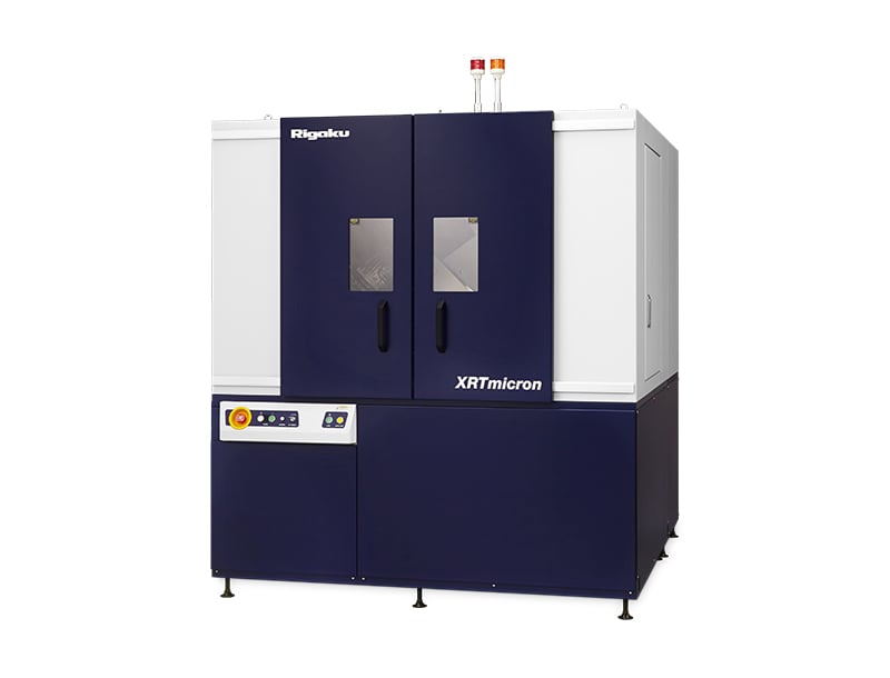
XRTmicron (Lab)
XRT (X-ray topography) imaging system for emerging power electronics, optoelectronics, and 5G / 6G communications (SiC, GaN, GaAs, InP, Ga₂O₃) for up to 300 mm wafers
Learn more
SmartLab
Advanced, state-of-the-art, high-resolution XRD system powered by Guidance expert system software
Learn more
XtaLAB Synergy-ED
A new and fully integrated electron diffractometer for measuring submicron crystals, utilizing a seamless workflow from data collection to structure determination of crystal structures.
Learn more
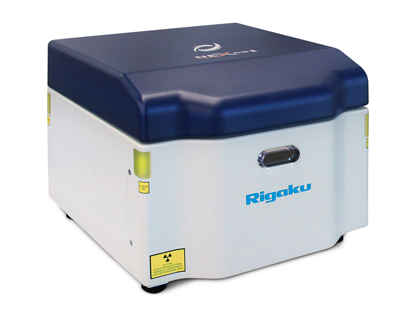
NEX CG II Series
High-performance indirect excitation EDXRF for complex applications with trace elements and variable base matrices
Learn more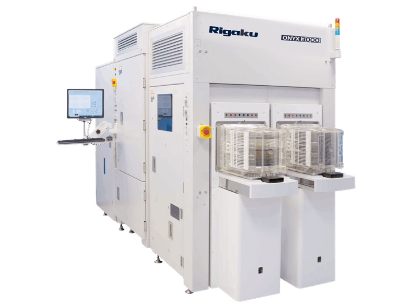
ONYX 3000
EDXRF and optical hybrid metrology tool for automated X-ray analysis, 3D scanning, and 2D microscope for film thickness and composition measurements on blanket and patterned wafers for up to 300 mm wafers
Learn more
Contact Us
Whether you're interested in getting a quote, want a demo, need technical support, or simply have a question, we're here to help.

Subscribe to a Rigaku Newsletter
Stay up to date with the latest Rigaku news!