Biotechnology & Life Sciences
From protein crystallography to molecular imaging
The scale of analysis used in biotechnology and life science research spans from the atomic level to the entirety of organisms. The X‑ray diffraction (XRD) technique has been widely used in pharmaceutical and food industries to study crystal structures at the atomic level and identify polymorphs. At the greater scale, we also offer preclinical multi-modal molecular imaging systems. For process control, X‑ray fluorescence (XRF) spectrometers and nondestructive testing (NDT) with radiography are used to detect heavy metals and foreign objects. Raman handheld analyzers are also used for raw material identification (RMID). Rigaku's analytical instruments are optimized for each scale and analytical purposes to keep their operation simple.

Related industries
Select your area of research or industry and explore more.
Pharmaceuticals
XRD and XRF are vital in drug discovery and quality control. X-ray and electron diffraction techniques are used for macromolecular and small molecule structure determination.
Learn more >

Life science research
X-ray and electron diffractometers play a critical role in life science by solving protein and small molecule structures at the early stages of research and development. We also offer multi-modal molecular imaging for pre-clinical studies and X-ray CT for soft tissue and plant imaging.
Learn more >

Biotechnology
XRD and crystallography contribute to crystal structure studies in the biotechnology industry. SAXS is applied to nanometer-scale periodic structures. Meanwhile, XRF is often used for elemental analysis. X-ray CT is also used to study both naturally occurring and engineered biological materials.
Learn more >

Food and food ingredients
On the process control side, at-line monitoring, such as XRF for heavy metal concentration analysis and NDT screening for foreign objects, can enhance cost efficiency. On the research and development side, XRD and SAXS are excellent tools for studying food ingredient structures at the molecular level.
Learn more >

Cosmetics
You can use XRD and XRF for structural and elemental analyses both in the research and process control stages. Handheld Raman analyzers can analyze materials through packaging and are used for RMID, verification, and authentication for anti-counterfeit prevention.
Learn more >Related products
Many products contribute to the biotechnology and life science research and process control. Use the Product Finder to find the best match for your needs.
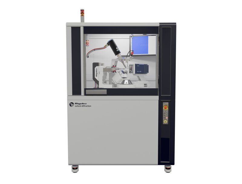
XtaLAB Synergy-S
Our most popular diffractometer for Chemical Crystallography and Mineralogy, configured with either single or dual microfocus sealed tube X-ray sources and an extremely low noise direct X-ray detection detector.
Learn moreXtaLAB Synergy-R
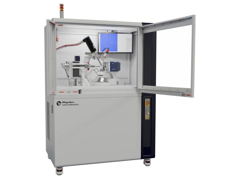
Our most popular diffractometer for Protein Crystallography, configured with a high-flux rotating anode X-ray source and an extremely low noise direct X-ray detection detector.
Learn moreXtaLAB Synergy-ED
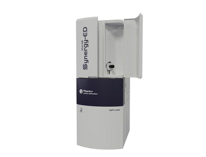
A new and fully integrated electron diffractometer for measuring submicron crystals, utilizing a seamless workflow from data collection to structure determination of crystal structures.
Learn moreSmartLab
Advanced, state-of-the-art, high-resolution XRD system powered by Guidance expert system software
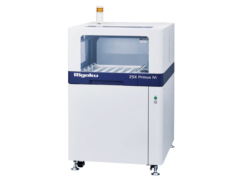
Learn moreZSX Primus IV𝒾
High-power, tube-below, sequential WDXRF spectrometer with new ZSX Guidance expert system software
Learn moreMILabs - a Rigaku Company Molecular imaging
MILabs is one of the global leaders in molecular imaging and offers high-performance pre-clinical multi-modal imaging systems that can integrate micro-PET, micro-SPECT, micro-CT, and Optical Tomography systems.
Application notes
Explore the example analyses to see which analytical technique is right for you.
-
XRD1003 - How to Evaluate Solid Pharmaceutical Drugs (3): Confirming Hydrates
-
XRD1002 - How to Evaluate Solid Pharmaceutical Drugs (2): Confirming the Presence/absence of Amorphous Substances
-
XRD1001 - How to Evaluate Solid Pharmaceutical Drugs (1): Confirming the crystal form of an API
-
XCT2201 - Submicron CT for the Life Sciences: Animal and Tissue Research Applications
-
XCT2103 - Submicron CT for Plant Research Applications
-
XCT2102 - Submicron CT for the Pharmaceutical Industry
-
WDXRF1102 - Standardless FP Analysis of Plant, Animal and Food Samples Applying Correction by Scattering Line
-
TA6017 - Evolved Gas and Kinetic Analysis of Acetylsalicylic Acid
-
TA6014 - Evaluation of Chocolates
-
TA6006 - Thermal Behavior Analysis of Over-the-counter (OTC) Drugs
-
TA6005 - Comparison of Drugs
-
TA5012 - Melting of an Extremely Small Amount of Dotriacontane
-
TA-TGMS-Pharm - TG-MS Analysis for Thermal Behavior of Commercialized PharmaceuticalProducts
-
SMX042 - In situ crystallization and diffraction experiments with the Insight Chips liquid nano-channel holder and XtaLAB Synergy-ED
-
SMX039 - The Absolute Structure of Light-atom-containing Molecules
-
SMX037 - Identification of a Fungicide Degradant using the XtaLAB Synergy-ED
-
SMX030 - Application of the Crystalline Sponge Method for Structure Determination of Persistent organic Pollutants of Agrochemical Degradation Products
-
SMX027 - Micro Powder Diffraction on XtaLAB Synergy Single Crystal Diffractometers
-
SMX010 - A Study of the Stereochemistry of Sucrose and a Phenidate Derivative using the Copper Microfocus Source of the XtaLAB Synergy-i
-
RAD006 - Pharmaceutical Excipients Identification with Handheld Raman
-
RAD002 - Regulation of Raw Material Identification in Cosmetics
-
RACCT9046 - Wild Bird Forensic
-
RACCT9045 - Tablet Crystallinity Analysis by X-ray CT
-
RACCT9044 - Rapid-release Pain Medication Capsule Comparison by X-ray CT
-
RACCT9043 - Multivitamin Tablet Analysis by X-ray CT
-
RACCT9042 - Microparticle Coating Analysis by X-ray CT
-
RACCT9041 - Famotidine Tablet Comparison by X-ray CT
-
RACCT9040 - Degradation of Sustained-release Dosage Tablet Imaged by X-ray CT
-
RACCT9039 - Compression Methods Comparison by X-ray CT
-
RACCT9038 - Brand Name vs Generic Atorvastatin Tablets Comparison by X-ray CT
-
RACCT9037 - Aspirin Tablet Coating Delamination Imaging by X-ray CT
-
RACCT9024 - Tomato Plant Root System Analysis by X-ray CT
-
RACCT9023 - Maize Kernel Analysis by X-ray CT
-
RACCT9022 - Coral Surface Area and Porosity Analysis by X-ray CT
-
RACCT9021 - Cicada Analysis by X-ray CT
-
RACCT9012 - Processed Meat Heating Experiment by X-ray CT
-
RACCT9011 - Processed Meat Fat and Protein Analysis Distribution Analysis by X-ray CT
-
RACCT9010 - Pretzel Coating Thickness Analysis by X-ray CT
-
RACCT9009 - Chocolate Candy Coating Analysis by X-ray CT
-
RACCT9008 - Breading Air and Oil Distribution Analysis by X-ray CT
-
RACCT9007 - Bread Dough Rising Process Observed by X-ray CT
-
RACCT9003 - Makeup Sponge Wear and Tear Analysis by X-ray CT
-
PX031 - Method: MicroED/3D ED on Proteins Using the XtaLAB Synergy-ED
-
PX030 - Using CrysAlisᴾʳᵒ to Tackle Macromolecular Non-merohedral Twins
-
PX029 - Intoducing the XtaLAB Synergy Flow
-
PX028 - Resolving Large Unit Cells by using the Correct Detector, Distance, and Beam Divergence
-
PX027 - Synergy Of PX and SMX on One Diffractometer with the HyPix-Arc 150°
-
PX026 - Sulfur SAD Phasing of Thaumatin in 10 Minutes with the HyPix-Arc 150° and XtaLAB Synergy-DW
-
PX024 - Lysozyme S-SAD Phasing with Two Minutes of Data Collection on the XtaLAB SynergyCustom
-
PX023 - ClpS SAD phasing with 30 Minutes of Data Collection on the XtaLAB Synergy-S
-
PX022 - Thaumatin S-SAD Phasing with 2 hours of Data Collection on the XtaLAB Synergy-S
-
PX021 - I-SAD Phasing of the HIV Integrase Core Domain using Data Collected on the XtaLAB Synergy-S
-
PX020 - In situ Data Collection and Structure Solution using the XtalCheck-S System
-
PX019 - Using the EIGER 4M for Single Crystal X-ray Diffraction on Catalase
-
PX018 - CrysAlisᴾʳᵒ: An All-in-one Software Package for Single Crystal X-ray Diffraction
-
PX015 - Data Collection and Improved Spot Resolution with the HyPix-6000HE HPC Detector
-
PHRM0002 - In-Situ DSC-Humidity PXRD Analysis for Pharmaceuticals
-
PHRM0001 - Crystalline Sponge Method on XtaLAB Synergy Systems
-
EDXRF3226 - NEX CG II - Analysis of Nutrients in Silage
-
EDXRF3198 - NEX CG II - Animal Feed Analysis
-
EDXRF3186 - NEX DE - Nutrient Premixes for Milled Grain Enrichment
-
EDXRF1933 - NEX DE Series—Catalysts Residue and Heavy Metal Screening in Pharma Formulation Process Chemistry
-
EDXRF1877 - NEX QC+—TiO₂ and ZnO in Lotions
-
EDXRF1858 - NEX QC+ QuantEZ—P₂O₅ and K₂O in Fertilizers
-
EDXRF1552 - NEX QC+—Nutrients in Grass Silage
-
EDXRF1491 - NEX QC+—Iron in Milk Powder
-
EDXRF1425 - NEX QC—Analysis of Iron in Straw
-
EDXRF1420 - NEX QC+—Chlorine and Ash in Biomass
-
EDXRF1385 - Agricultural Soils and Plant Materials
-
B-XRI1023 - Analysis of potato thickness and oil particle size in potato chips by micro X-ray CT
-
B-XRI1017 - Analysis of volume fraction and thickness of bran layer using a high-resolution 3D X-ray microscope
-
B-XRI1015 - Observation of Lyophilized Formulations by Micro X-ray CT
-
B-XRI1002 - Observation of Milk Protein Aggregation in String Cheese
-
B-XRD1142 - Quantitative Analysis of Crystal Polymorphs by the DD Method
-
B-XRD1137 - Phase ID Analysis of Micro-impurities on the Surface of a Tablet by Micro-area XRD Measurements
-
B-XRD1135 - Observation of Butter Crystal by Simultaneous XRD-DSC Measurement
-
B-XRD1133 - Observation of Phase Transition Behavior of Pharmaceutical Materials with a Benchtop XRD System
-
B-XRD1129 - Variable Humidity Measurement of a Drug Substance using XRD-DSC and a Humidity Controller
-
B-XRD1128 - Observation of Dehydration Behavior of a Drug Substance using TG-DTA and XRD-DSC
-
B-XRD1126 - Crystallization of Chocolate Observed by XRD-DSC
-
B-XRD1125 - Quantification of the Taste of Salt by DD (Direct Derivation) Method
-
B-XRD1120 - Quantitative Analysis of Pharmaceutical Polymorphic Forms via the DD Method
-
B-XRD1114 - LOQ of Trace Impurities in API by the DD Method
-
B-XRD1108 - Direct Observation of Melting and Crystallization of Fresh Cream
-
B-XRD1081 - Quantitative Analysis of Polymorphic Impurities in a Drug Substance by the Calibration Method
-
B-XRD1060 - MiniFlex Measurement of Trace Samples
-
B-XRD1046 - Crystal Phase Identification of Pharmaceutical Materials in the Freeze Drying Process
-
B-XRD1043 - Powder Crystal Structure of Organic Crystals
-
B-XRD1035 - Crystal Structure Analysis of a Powder Sample of Pharmaceutical Cocrystals by the Rietveld Method
-
B-XRD1023 - Measurement of Pseudo-polymorph Impurities in Tablets
-
B-XRD1021 - Phase Changes of Pharmaceuticals as a Function of Temperature and Humidity
-
B-XRD1020 - Observation of Dehydration Process of Hydrate by XRD-DSC
-
B-TA2042 - Thermal Stability of Polyethylene Glycol Studied by TG-MS
-
B-TA2025 - Evolved Gas Analysis in Edible Oil by STA-MS
-
B-TA1086 - Analysis of Polymorphic Transition of Carbamazepine -Sample Observation DSC, ChromTA (Chromatic Thermal Analysis)
-
B-TA1058 - Thermal Decomposition Behavior of Edible Oils: Effects of Degradation
-
B-TA1050 - Gelatinization of Starch in Rice and Wheat Flour
-
B-TA1044 - Vaporization rate of H₂O below room temperature
-
B-TA1040 - Thermal analysis of gummy candy -Part 2-~Sample observation DSC~
-
B-TA1037 - Thermal Behavior of Aspartame by Sample Observation STA
-
B-TA1035 - Dehydration of Trehalose Dihydrate by STA
-
B-TA1030 - Thermal Decomposition of Starch-based Filler by Sample Observation STA
-
B-TA1027 - Water Absorption Behavior of Materials for Biomedical Application by HUM-TG
-
B-TA1020 - Characterization of Olive Oil by DSC
-
B-TA1019 - Thermal Analysis of Red Palm Oil by STA
-
B-TA1017 - Thermal Analysis of Pure Cocoa by DSC
-
B-TA1013 - Mechanically Induced Amorphization in Trehalose Dihydrate by STA
-
B-TA1011 - Artificial Sweetener and Table Sugar by STA
-
B-TA1010 - Thermal Behavior of Caffeine by STA and DSC
-
B-TA1008 - Mechanochemical Effect in Caffeine
-
B-TA1003 - The Behavior of Water Absorption and Dehydration of Organic Materials
-
B-SCX1011 - High-Speed Measurement Of A Peptide Drug Using HyPix-Arc 150°
Contact Us
Whether you're interested in getting a quote, want a demo, need technical support, or simply have a question, we're here to help.

Subscribe to a Rigaku Newsletter
Stay up to date with the latest Rigaku news!