Application Note B-XRI1017
Introduction
The internal structure of plants is commonly observed and measured by optical microscopes. For example, in the quality evaluation of paddy rice, the size of brown rice and the thickness of the bran layer are measured. These measurements require preprocessing, such as ethanol soaking, staining, and cutting. 3D X-ray microscopy (X-ray CT) enables 3D imaging of samples without preprocessing, which allows the original structure of plants to be captured by nondestructive observation. A paddy rice grain was scanned using a 3D X-ray microscope. Then, the volume fraction and thickness distribution of the bran layer were calculated.
Measurement and analysis
A paddy rice grain (Figure 1) was scanned with a Mo source at 5.1 μm/voxel for ten minutes to produce 3D rendered images (Figure 2). The bran layer (the layer on the surface of the brown rice), the endosperm (white rice inside the brown rice), and the embryo (protrusion at the tip of the brown rice) were observed in the CT cross-section image. The bran layer, which contains iron and magnesium, appeared whiter than the endosperm because of the difference in X-ray transmittance. The image voxels were segmented into three phases based on the gray values.
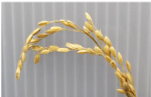
Figure 1: Paddy rice grains
 Figure 2: 3D rendered images of a paddy rice grain
Next, focusing on the bran layer, the volume fractions of the bran layer (excluding the embryo) in brown rice (excluding the hull but including the embryo) were calculated (Table 1). In addition, the thickness of the bran layer was color-coded in the 3D rendered image (Figure 3). The image shows the three-dimensional distribution of the bran layer. For example, the bran layer thickness differs between the “belly” and “back” of the brown rice.
Table 1: Volume fraction of the bran layer
| Volume of brown rice | 18.5 mm³ |
| Volume of bran layer/td> | 1.1 mm³ |
| Volume fraction of bran layer | 5.9 vol% |

Figure 3: 3D rendered image of the thickness distribution of the bran layer
Recommended equipment and software
