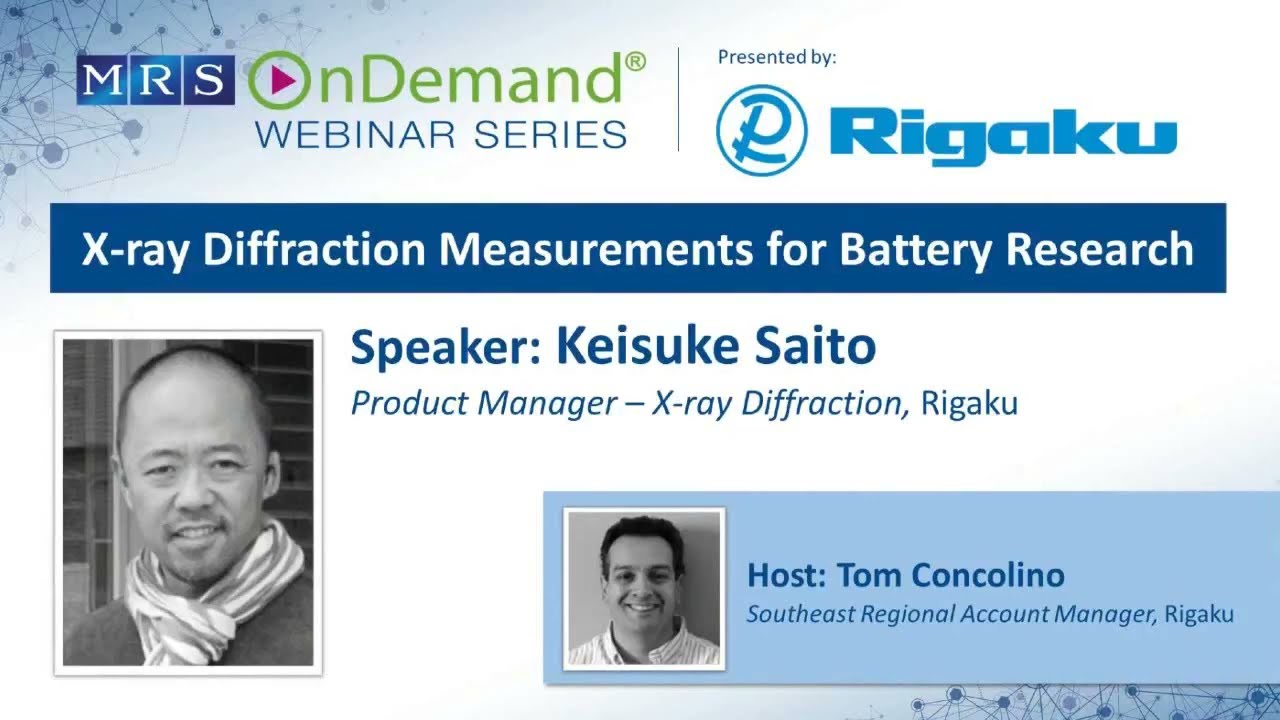
X-ray Diffraction Measurements for Battery Research

This is a written summary of a live webinar presented on March 21, 2024. The recording and resources are available on the recording page.
Presented by:

Webinar summary
The webinar offers a detailed and practical overview of how modern X-ray diffraction (XRD) and scattering techniques are applied to the study of battery materials, particularly lithium-ion and solid-state batteries. The session is highly relevant to battery researchers aiming to understand structural transformations during charge-discharge cycles, and how operando measurements and complementary techniques can unlock deeper insights into material performance and degradation.
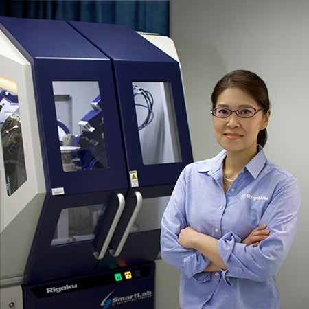
Keisuke begins by framing XRD as an indispensable tool for analyzing both the crystal and local structures of materials. He introduces two major instrument classes: the benchtop MiniFlex for routine crystal structure analysis and the floor-standing SmartLab, which supports advanced capabilities like operando measurements and local/global structural analysis. The SmartLab system features modular hardware, fully motorized optical alignment, and intuitive software that automatically configures instrument settings based on the selected application—making it highly adaptable for complex in situ experiments. A standout feature is the HyPix 3000, a 2D silicon-based detector with excellent energy resolution and a wide 2θ capture range, allowing for fast, real-time data acquisition without scanning the goniometer. This makes it especially powerful for studying dynamic processes such as phase transformations during electrochemical cycling or heating.
Several case studies are presented to highlight the application of XRD in battery research. For example, operando measurements on LiNiMnCo (NMC) oxide cathodes show clear phase transformations as lithium ions move in and out of the structure. Shifts in key diffraction peaks (e.g., 003 and 101) during cycling are used to infer changes in lattice parameters, with the c-axis expanding during charging and the a and b axes contracting—trends which reverse upon discharge. Such detailed tracking of lattice behavior is vital to understanding capacity fade and mechanical stress development in layered oxide materials.
In studies involving carbon anodes, the formation of different lithiated graphite phases (LiC₆, LiC₁₂, LiC₃₆, and LiC₇₂) is resolved via XRD. The technique reveals distinct stage transitions during lithiation and delithiation, with some stages—like stage 3—barely distinguishable despite the compression effect of high-energy X-rays thanks to the instrument's high-resolution. Here, SmartLab's ability to easily switch X-ray sources (e.g., from copper to molybdenum or silver) allows researchers to optimize resolution and penetration depth for their specific sample geometry and composition.
The webinar also delves into high-rate capability measurements using lithium iron phosphate (LFP). By analyzing changes in peak intensities under different C-rates, the speaker demonstrates how XRD can diagnose whether lithium-ion diffusion is kinetically limited, as seen at higher rates like 5C. For materials development, such insights are crucial for tuning particle morphology and optimizing electrode architecture.
Another advanced topic covered is crystallite size determination. Rigaku employs the Fundamental Parameters (FP) method, which calculates the instrument profile function without requiring a reference standard. This method allows for accurate estimation of crystallite sizes up to 200 nm—surpassing the traditional Scherrer formula. Differences in crystallite size distribution between LFP samples are used to explain variations in diffraction peak sharpness and tailing. The FP method supports various statistical distribution models (e.g., gamma, log-normal), and researchers are encouraged to base their choice on complementary methods such as TEM imaging.
The bond valence sum (BVS) analysis, derived from Rietveld refinement, is presented as a powerful tool to map lithium-ion conduction paths within a unit cell. Using this approach, researchers can identify regions where Li⁺ ions are most stable and likely to migrate, which is instrumental in the design of high-conductivity solid-state electrolytes.
Perhaps the most advanced segment of the webinar is the discussion of pair distribution function (PDF) analysis. By combining Bragg and diffuse scattering through total scattering measurements, PDF reveals both long-range and short-range atomic order. This is particularly important for materials that are amorphous, nanocrystalline, or structurally disordered. For example, in a study of LiMn₂O₄, Rietveld refinement alone could not distinguish between cubic and orthorhombic phases, whereas PDF analysis clearly favored an orthorhombic local structure—consistent with observed insulating behavior despite a nominally metallic cubic symmetry. The example underscores how PDF can complement conventional XRD and resolve ambiguities in structure-property correlations.
Overall, the webinar demonstrates how Rigaku’s SmartLab system, with its flexible geometry, advanced detectors, and automation, is uniquely suited for high-throughput, high-precision X-ray analysis in battery research. Researchers are shown how to characterize phase transitions, lattice strain, ion transport pathways, crystallite size, and structural disorder—all under operando conditions. This holistic approach makes it possible to link atomic-scale changes to electrochemical performance, guiding the development of more efficient and durable battery materials.
Key questions answered in the webinar
-
X-ray diffraction (XRD) is a powerful analytical technique that examines both crystalline and local structures of materials. In battery research, XRD is widely used to analyze cathode, separator, solid electrolyte, and anode materials. Key applications include:
- Phase identification and quantification: Determining the different chemical phases present in a material and their relative amounts.
- Lattice parameters and crystal structure refinement: Precisely measuring the dimensions of the unit cell and refining the atomic positions within the crystal structure.
- Bond valence sum (BVS) application: Identifying potential ion conduction paths within a material, which is crucial for understanding how ions move during charging and discharging.
- Percent crystallinity: Quantifying the amorphous content in a material.
- Crystallite size measurement: Evaluating the size of the small single-crystal components within a material, which can impact performance.
- Operando measurements: Studying structural changes in battery components in real-time under working conditions (charging/discharging).
- Local and global structure analysis by pair distribution function (PDF): Analyzing both the short-range and long-range atomic order, providing a comprehensive understanding of the material's structure.
-
Operando measurements involve real-time observation of structural changes in battery materials while the battery is actively cycling (charging and discharging). This provides critical insights into the dynamic processes occurring within the battery. The SmartLab diffractometer supports various sample holders for operando measurements, including:
- Coin cells: Small, button-shaped cells often used for preliminary battery research.
- Electrochemical cells: Designed for both liquid-based and solid-based battery structures, allowing for controlled application of current and voltage.
- Pouch cells: Flexible, rectangular battery cells, with or without spatial mapping capabilities for detailed analysis of specific regions.
These setups can be integrated with a potentiostat (like Biologic SP 150 and VSP models) to synchronize the XRD measurements with the electrochemical cycling, enabling the correlation of structural changes with battery performance. Different X-ray geometries (refraction and transmission) and X-ray energies (e.g., copper, molybdenum, silver radiation) are used depending on the sample type and desired information.
-
Crystallite size analysis in XRD focuses on determining the size of the individual crystalline domains within a material, which are distinct from particle size (a particle can be composed of multiple crystallites). The SmartLab diffractometer utilizes a "fundamental parameters approach" (FP method) to calculate instrumental profile functions mathematically, allowing for precise determination of crystallite sizes ranging from 1 nanometer to 200 nanometers. This method is more accurate than the traditional Scherrer formula, which is limited to smaller crystallite sizes (up to 80-100 nm).
For cathode materials, understanding crystallite size is increasingly important as it significantly influences battery performance parameters like reaction kinetics and ion diffusion. For instance, smaller crystallites can offer more surface area for electrochemical reactions, potentially improving charge/discharge rates. By analyzing the broadening and shape of XRD peaks, researchers can gain insights into the crystallite size distribution within battery components.
-
The Bond Valence Sum (BVS) method is used in conjunction with Rietveld refinement to identify areas within a crystal structure where a specific atom is likely to maintain a particular oxidation state. After performing Rietveld refinement to determine the precise lattice parameters and atomic coordinates of a crystal, BVS calculations can map out regions where mobile ions (like lithium ions in batteries) are most likely to reside and move.
By highlighting these "bands" or pathways, BVS helps researchers visualize and understand the potential ion conduction paths within the material. This is crucial for designing new materials with improved ionic conductivity, which directly impacts the battery's power capability and charge/discharge efficiency. The method can be applied to identify how these paths change depending on the charging and discharging state of the battery.
-
Pair Distribution Function (PDF) analysis is a technique that provides both short-range (local) and long-range (global) structural information about materials. It is derived from the "total scattering" data, which includes both Bragg scattering (revealing long-range order and crystal structure) and diffuse scattering (revealing short-range atomic arrangements).
Unlike conventional XRD that primarily focuses on long-range order, PDF analysis allows researchers to study amorphous materials, semi-crystalline materials, and crystalline materials with local structural variations. By transforming the total scattering data, PDF generates a plot of atomic probability against interatomic distance. This enables the analysis of how atoms are arranged not just in the perfect crystalline lattice, but also in disordered or nanocrystalline regions. High-energy X-rays (like molybdenum or silver radiation) and specialized optics and detectors are recommended for effective PDF measurements due to the weak nature of diffuse scattering. PDF is complementary to Rietveld refinement, as it can reveal local structural details that might be averaged out or not discernible in standard Bragg diffraction patterns, such as distinguishing between slightly different crystal symmetries at a local level.
-
The SmartLab diffractometer supports a variety of X-ray sources, allowing researchers to choose the most appropriate radiation for their specific application:
- Copper (Cu): This is a conventional X-ray source often used for standard powder diffraction measurements, especially in refraction geometry for liquid-based battery cells.
- Molybdenum (Mo): A higher-energy X-ray source recommended for transmission geometry experiments and total scattering measurements (for PDF analysis) due to its better penetration through samples and wider Q-range coverage in reciprocal space.
- Silver (Ag): An even higher-energy X-ray source, also suitable for transmission geometry and PDF analysis, offering further advantages in penetration and Q-range coverage compared to molybdenum.
- Chromium (Cr) and cobalt (Co): These are typically used for specific applications like geological studies and are generally not employed for lithium-ion battery research.
The choice of X-ray source depends on the sample's characteristics (e.g., density, thickness), the desired geometry (refraction vs. transmission), and the type of information being sought (e.g., conventional crystal structure vs. local structure via PDF). The SmartLab system is designed to easily switch X-ray tubes, with automated optic alignment, making the transition between different radiations efficient.
-
The SmartLab diffractometer is equipped with user guidance software that significantly simplifies the setup and operation of XRD experiments. This software features predefined application packages, allowing users to simply select the desired experiment type (e.g., transmission SAXS measurement, operando in-situ measurement).
Upon selection, the software guides the user through the necessary steps, including:
- Optic configuration: Recommending and guiding the setup of the ideal detector, optics, and sample stage for the chosen application.
- Component exchange prompts: Displaying "smart messages" that inform the user which components (e.g., specific slits, sample stages, vacuum beam paths) need to be switched or exchanged.
- Automated alignment: Once components are configured, the software automatically aligns the X-ray source, optics, slits, sample stages, and detector. This includes motorized movements and precise positioning, with a complete alignment typically taking a maximum of 10 minutes. This automated process minimizes potential misalignments and ensures accurate and reproducible results, allowing users to focus more on their research.
-
The SmartLab diffractometer offers advanced detector options, such as the HyPix-3000, a two-dimensional silicon-based detector with several unique features that enhance XRD measurements, especially for battery research:
- Superior energy resolution (15%): This allows for effective suppression of X-ray fluorescence (XRF) signals from samples containing transition metals (like cobalt, manganese, nickel, iron), which can otherwise increase background noise in XRD data.
- Quick readout time: The detector covers a 30° 2θ range simultaneously, eliminating the need for goniometer scanning during in-situ operando measurements. This enables rapid data acquisition, allowing for X-ray diffractograms to be collected every second, crucial for observing fast dynamic processes.
- Optimized for Both High and Soft Energy X-rays: The HyPix-3000 can effectively detect a wide range of X-ray energies, from conventional copper radiation to higher energies required for techniques like Pair Distribution Function (PDF) analysis. This versatility means a single detector can be used for multiple applications without needing to be physically switched when changing X-ray sources.
- Multi-dimensional detection modes: While a 2D detector, the HyPix-3000 can simulate 0D (point detector) and 1D detection modes automatically based on the selected application (e.g., rocking curve, high-speed powder XRD), further expanding its versatility.

Subscribe to the Bridge newsletter
Stay up to date with materials analysis news and upcoming conferences, webinars and podcasts, as well as learning new analytical techniques and applications.

Contact Us
Whether you're interested in getting a quote, want a demo, need technical support, or simply have a question, we're here to help.
