XtaLAB Synergy-ED
Fully Integrated Electron Diffractometer
A system any X-ray crystallographer will find intuitive to operate
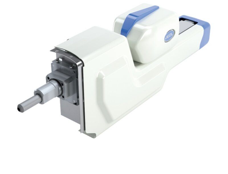
XtaLAB Synergy-ED is a new and fully integrated electron diffractometer, creating a seamless workflow from data collection to structure determination of three-dimensional molecular structures. The key feature of this product is that it provides researchers an integrated platform enabling easy access to electron crystallography. The XtaLAB Synergy-ED is a system any X-ray crystallographer will find intuitive to operate without having to become an expert in electron microscopy.
.jpg?width=800&height=610&name=XtaLAB%20Synergy-ED%20electron%20diffractometer%20partial%20cutaway).jpg)
.jpg?width=800&height=610&name=XtaLAB%20Synergy-ED%20electron%20diffractometer%20(top%20door%20open).jpg)
.jpg?width=800&height=610&name=XtaLAB%20Synergy-ED%20electron%20diffractometer%20(doors%20closed).jpg)
.jpg?width=800&height=610&name=XtaLAB%20Synergy-ED%20electron%20diffractometer%20(full%20cutaway).jpg)
XtaLAB Synergy-ED Overview
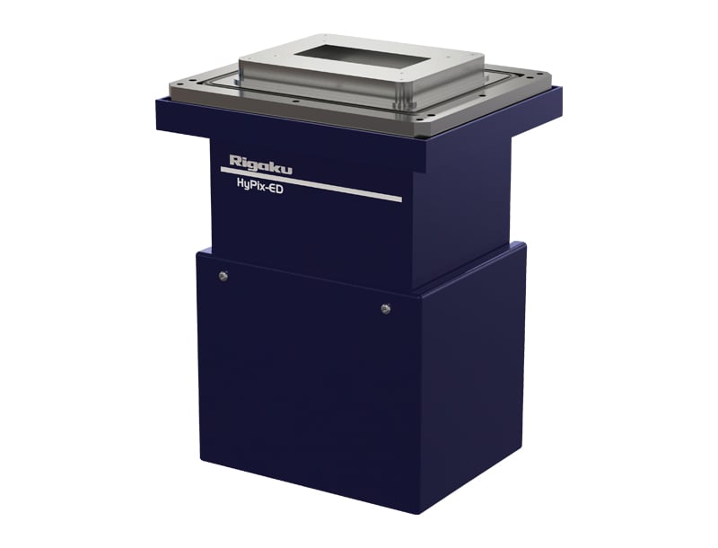
The XtaLAB Synergy-ED is the result of an innovative collaboration between Rigaku and JEOL, to synergistically combine our core technologies: Rigaku’s high-speed, high-sensitivity photon-counting detector (HyPix-ED) and state-of-the-art instrument control and single crystal analysis software platform (CrysAlisPro for ED), and JEOL’s long-term expertise and market leadership in designing and producing transmission electron microscopes.
Solve the unsolvable
Simply put, MicroED, also known as 3D ED, offers the chance to solve 3D structures from samples that are too small for X-ray diffraction. For X-ray crystallography, these samples would yield insufficient diffraction strength or quality to obtain a fully resolved structure.
When you are faced with a sample that is tricky to crystallize, MicroED can let you shortcut the difficult process of optimizing the crystallization conditions. Instead, the technique allows you to obtain 3D data and structure from powders, whether you have a lot or just a little. The only requirements are that you have crystalline material and that the grains are below 1 micron in size.
It doesn’t matter if you are studying pharmaceuticals, MOFs or any other materials. If you are struggling to grow crystals, the XtaLAB Synergy-ED may help you solve your unsolvable problems.
The value of dedicated instrumentation
The XtaLAB Synergy-ED is a dedicated electron diffractometer that is operated by the same CrysAlisPro control software used to run Rigaku’s X-ray diffractometers. Those who have used TEMs for diffraction experiments know the pain of sharing time with microscopists and/or configuring the hardware before and after use. XtaLAB Synergy-ED offers you a device specifically for diffraction so it is always calibrated and ready for diffraction and can even be installed alongside your X-ray instrumentation†. CrysAlisPro controls all aspects of the diffractometer, removing the need for specialist knowledge, so even if you’ve never used an electron diffractometer before, you’ll be solving structures in no time.
Powerful software
All of the powerful tools X-ray crystallographers have become accustomed to are available for XtaLAB Synergy-ED users as well. With CrysAlisPro-ED, you no longer need to chain together multiple pieces of software and figure out how to convert the data between each to get the job done. CrysAlisPro offers a seamless experience to get you all the way from selecting grains to measurement through to publishable data with one piece of software.
†please check with your local Rigaku representative for site requirements
XtaLAB Synergy-ED Features
XtaLAB Synergy-ED Videos
XtaLAB Synergy-ED Specifications
| Technique | 3D microcrystal electron diffraction (MicroED/3D ED) with continuous-rotation data collection | |
|---|---|---|
| Attributes | Dedicated electron diffractometer running CrysAlisPro-ED, a complete integrated software platform. Ultra-low-dose operation for sample screening and diffraction data collection. Optimized electron optics for versatile diffraction conditions without the need for regular alignment or calibration. Automation features for unattended data collection from hundreds of microcrystals | |
| Detector | High-speed, high-sensitivity hybrid pixel array detector, HyPix-ED. Variable detector distance via electron-optical projection system (10x range) | |
| Source | LaB₆ electron gun, electron energy up to 200 keV (λ = 0.0251 Å). Three-stage electron-optical condenser system | |
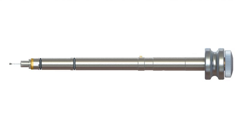
| Goniometer | Side-entry, single rotation axis with 160° total range | |
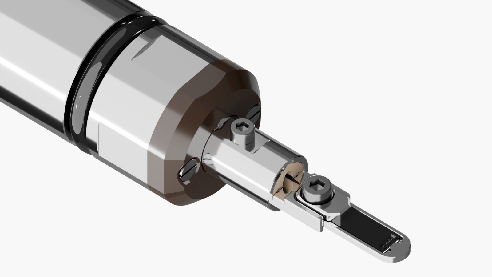
| Accessories | Cryo-transfer device, low-temperature, heating, gas-cell, and inert-transfer sample holders. Elemental analysis (EDS/EDX) | |
| Computer | External PC, MS Windows OS | |
XtaLAB Synergy-ED Options
The following accessories are available for this product:
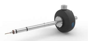
Cryo transfer holder from Simple Origin
A cryo-transfer sample holder offering the ability to load two standard 3 mm grids simultaneously
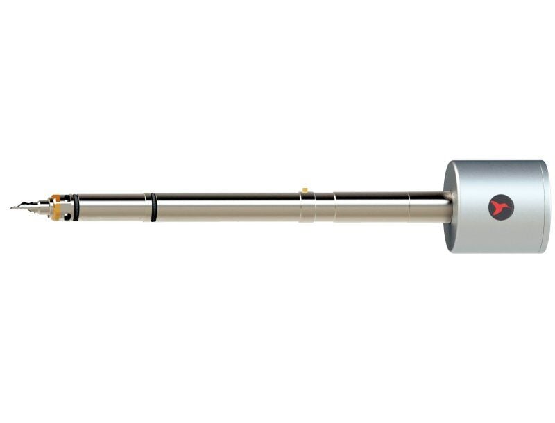
Air-free transfer sample holder from Hummingbird Scientific
Air-free transfer sample holder designed for transfer of air-sensitive samples from preparation environments without exposure to ambient conditions.
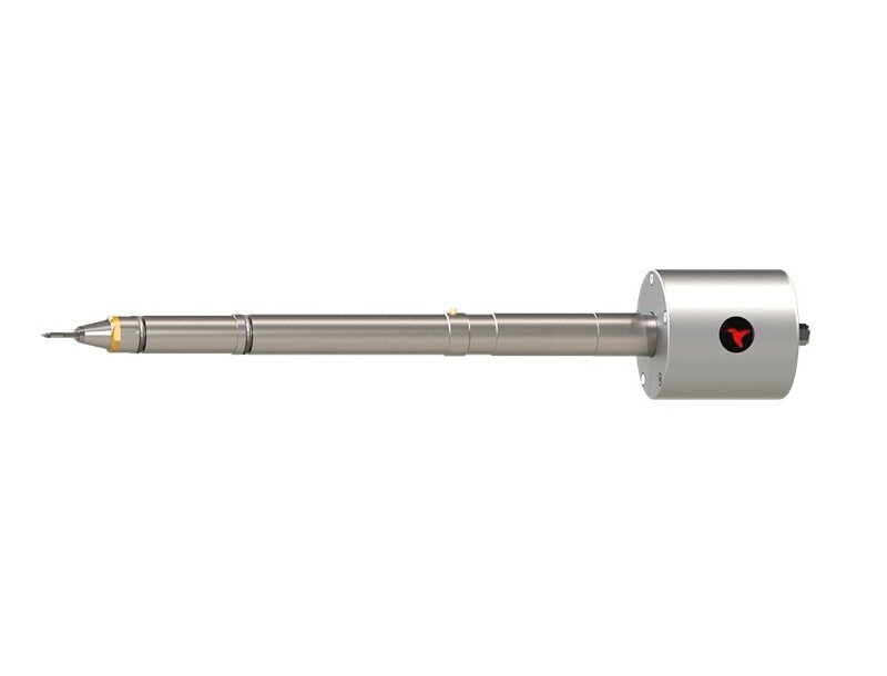
Insight Chips Liquid Nano-Channel Sample Holder
The Liquid Nano-Channel Sample Holder enables electron diffraction experiments in liquid environments with unmatched simplicity and precision.
X-ray spectrometer (JEOL JED-2300)
A fully integrated energy dispersive X-ray spectrometer providing elemental analysis on each single nanocrystal.
Cryo-transfer sample holder
Using this sample holder, a sample grid is cooled to liquid-nitrogen temperature prior to insertion, and transferred into vacuum in a protected dry environment.
High-temperature sample holder
This holder combines agile and accurate temperature control up to 1000°C with unmatched ease of use.
Gas-cell sample holder
The gas heating cell sample holder allows a specimen to be exposed to a gas environment and elevated temperatures, allowing for observation of the crystal structure during complex catalysis, adsorption, phase change, or growth processes.
XtaLAB Synergy-ED Application Notes
The following application notes are relevant to this product
-
SMX042 - In situ crystallization and diffraction experiments with the Insight Chips liquid nano-channel holder and XtaLAB Synergy-ED
-
PX031 - Method: MicroED/3D ED on Proteins Using the XtaLAB Synergy-ED
-
SMX041 - Observing a Temperature-induced Phase Transition of Cu(ta)₂, a MOF, with XtaLAB Synergy-ED
-
SMX033 - XtaLAB Synergy-ED: A True Electron Diffractometer
-
SMX037 - Identification of a Fungicide Degradant using the XtaLAB Synergy-ED
XtaLAB Synergy-ED Resources
White paper
| Read the White Paper |
Webinars
| Solving Pharma’s Toughest Solid Form Challenges with Electron Diffraction | Register |
| Revealing Atomic-Level Battery Material Structures with Electron Diffraction | Register |
| Decoding the Complex Structures of MOFs for Design & Performance Insights | Register |
| Simple Electron Diffraction Workflow from Sample Prep to Structural Solutions | Watch the Recording |
| The Transformative Potential of Electron Diffraction | Watch the Recording |
| How MicroED Is Reshaping Materials, Life Science, and Chemistry Research | Watch the Recording |
| Illuminating the World of Sub-micron Crystal Structures with the XtaLAB Synergy-ED: A Review | Watch the Recording |
| 3D-ED in Pharmaceutical Compounds: Can we Measure Everything? | Watch the Recording |
| XtaLAB Synergy-ED: Single Crystal Structures from Powders | Watch the Recording |
| XtaLAB Synergy-ED Progress and Latest Results | Watch the Recording |
| MicroED: An Update from the Rigaku/JEOL Collaboration | Watch the Recording |
Rigaku Journal articles
| Read the Article | |
| Read the Article | |
| Read the Article |
Publications
Visit the Publication Library to access articles relevant to XtaLAB Synergy-ED
Forums
XtaLAB Synergy-ED Events
Learn more about our products at these events
-
EventDatesLocationEvent website
-
Rigaku School for Practical CrystallographyJanuary 26 2026 - February 6 2026
-
34th Annual Meeting of the German Crystallographic Society (DGK)February 25 2026 - February 28 2026Lübeck, Germany
-
Pittcon 2026March 9 2026 - March 11 2026San Antonio, TX, USA
-
BCA Spring Meeting 2026March 30 2026 - April 1 2026Leeds, United Kingdom
-
27th Congress and General Assembly of the IUCrAugust 11 2026 - August 18 2026Calgary, Alberta, Canada
XtaLAB Synergy-ED
Contact Us
Whether you're interested in getting a quote, want a demo, need technical support, or simply have a question, we're here to help.

