CT Products
Research-grade CT scanners
Resolution, versatility, and measurement speed are all important elements. However, for most measurement techniques, they are also in a tradeoff relationship. For X‑ray CT (computed tomography) scanners or X‑ray microscopes, the best combination of the X‑ray source, geometry, and detector is different depending on the desired resolution, sample size, and measurement speed.
CT Lab HX for flexible benchtop CT
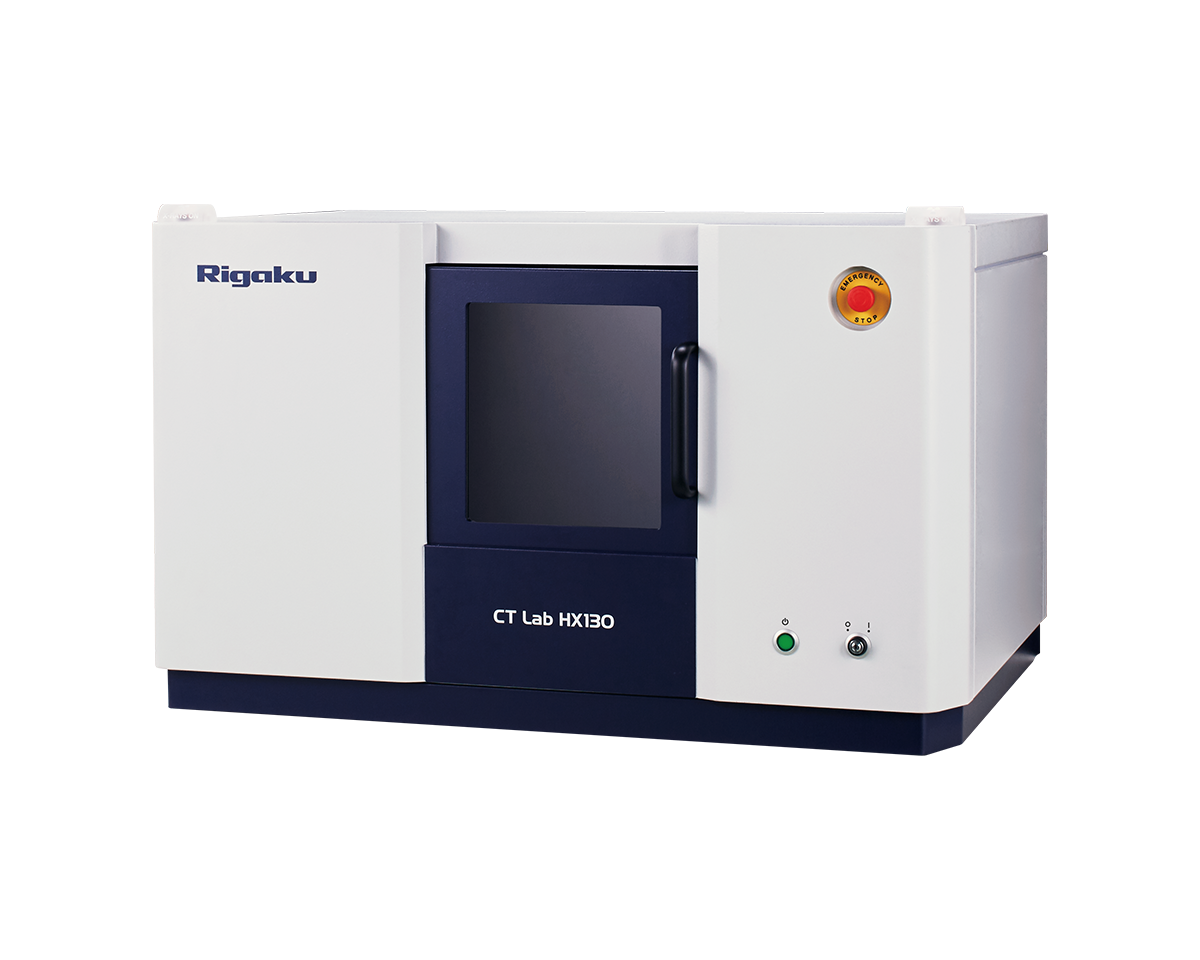
Rigaku CT Lab HX is a benchtop micro-CT scanner designed to accommodate a wide range of sample sizes, materials, and imaging requirements. Adjustable source-to-object distance (SOD) and source-to-detector distance (SDD) allow users to balance field of view and resolution for each application.
The system spans from a 2.1 µm voxel size in high-resolution mode to a 200 mm field of view in large-FOV mode. A 130 kV, 39 W X-ray source, along with adjustable X-ray conditions and filters, lets you optimize imaging for samples with different dimensions and attenuation.
This combination of adjustable geometry, energy, filtration, and field of view makes the CT Lab HX a flexible benchtop platform for routine research, failure analysis, and materials characterization.
CT Lab HR160 for accessible high-resolution 3D imaging
Rigaku CT Lab HR160 is a 160 kV high-resolution X-ray CT scanner designed for research, process development, and design engineering. The nanofocus transmission X-ray source and lens-free cone-beam geometry provide true 250 nm spatial resolution with a minimum voxel size of 62 nm.
The source-to-object distance (SOD) and source-to-detector distance (SDD) can be adjusted to optimize field of view and resolution without changing lenses, sources, or detectors. The 40–160 kV X-ray voltage range supports imaging of materials ranging from polymers and composites to electronics, batteries, and other engineered materials.
A streamlined software workflow guides users from scan setup through acquisition, reconstruction, and viewing, making high-resolution CT easier to configure and use without unnecessary hardware complexity.
CT Lab HV for large and dense industrial samples
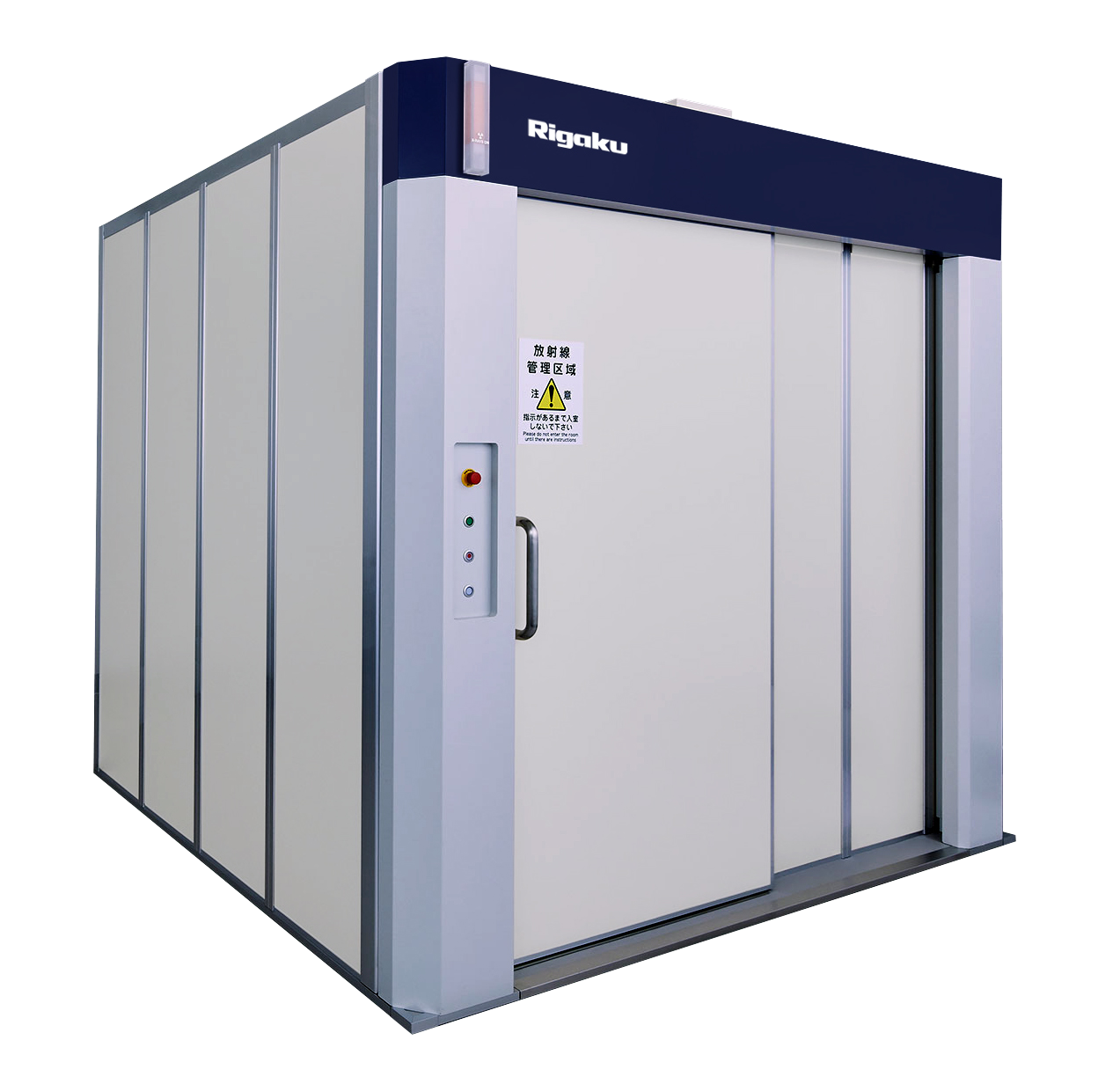
Rigaku CT Lab HV is a high-voltage industrial X-ray CT (computed tomography) scanner designed for non-destructive inspection of large and higher-density samples. Its large radiation enclosure, wide door opening, 225 kV X-ray source, and adjustable source-to-object and source-to-detector distances provide flexibility for a broad range of industrial components.
It accommodates samples up to 600 mm in diameter × 1200 mm in height, with a field of view up to 350 mm. For smaller regions requiring greater detail, the high-resolution configuration achieves true 3 µm spatial resolution.
These capabilities make the CT Lab HV well suited to industrial failure analysis, assembly inspection, dimensional evaluation, and characterization of dense or large components that are difficult to examine with lower-energy or smaller-volume CT systems.
nano3DX for resolution of low-density samples
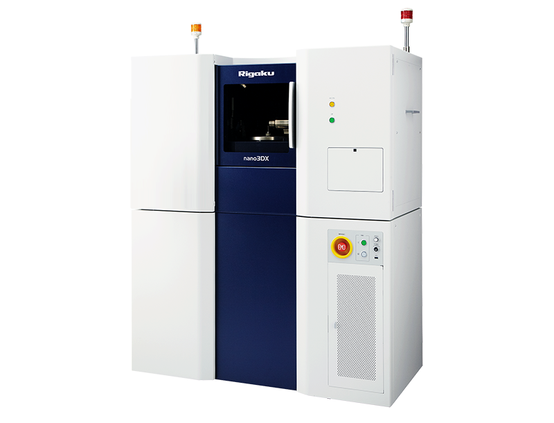
Rigaku nano3DX is a true submicron-resolution CT scanner designed for high-contrast imaging of low-density materials. Its parallel beam geometry and ultra-bright 1200 W rotating-anode X-ray source use low-energy, pseudo-monochromatic X-rays to improve contrast in materials that can be difficult to distinguish with higher-energy sources.
The X-ray anode can be selected from Cr (5.4 keV), Cu (8 keV), or Mo (17 keV), allowing the X-ray energy to match the sample material and size. With the highest-magnification lens, nano3DX achieves a 188 nm voxel size and true submicron spatial resolution of 500 nm.
CT Lab GX for fast and in-situ CT
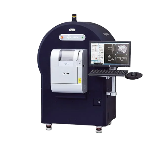
Rigaku CT Lab GX is a micro-CT scanner designed for fast imaging and experiments where the sample must remain stationary. Its gantry geometry rotates the X-ray source and detector around the sample, similar to a medical CT scanner, making it easier to mount irregular samples or connect samples to in-situ devices.
The combination of a high-power X-ray source, available in 90 kV/8 W or 130 kV/39 W configurations, and compact gantry geometry enables CT scans as fast as 3.9 seconds.
This high-speed capability supports time-resolved 4D CT, in-situ experiments, and rapid quality inspection where capturing structural changes over time or minimizing acquisition time is important.
Compare products
| CT Lab HX | CT Lab HR160 | CT Lab HV | nano3DX | CT Lab GX | |
| Minimum voxel resolution | 2.1 µm | 62 nm | 1.5 µm | 188 nm | 4.5 µm |
| Field of view (FOV) | 5 — 200 mm | 0.2 — 103 mm | 4 — 350 mm | 0.66 — 20 mm | 5 — 72 mm |
| Maximum sample size | ϕ200 mm x 270 mm height | ϕ250 mm x458 mm length | ϕ600 mm x 1200 mm in height | ϕ20 mm x 40 mm height | ϕ163 mm x 398 mm length |
| Geometry | Cone beam geometry | Cone beam geometry | Cone beam geometry | Parallel beam geometry | Cone beam gantry geometry |
| X-ray source | 39 W traditional microsource | 16.6 W traditional nanosource | 350 W microsource | 1200 W rotating anode microsource | 8 or 39 W traditional microsource |
| X-ray source applied voltage | 130 kV | 160 kV | 225 kV | 60 kV (Characteristic X-ray energies at 5.4, 8.0, and 17.4 keV) | 90 kV or 130 kV |
| X-ray detector | Flatpanel | Flatpanel | Flatpanel | High-resolution sCMOS | Flatpanel |
| Price | $$ | $$$ | $$$$ | $$$$ | $$$ |
| Dimensions | 980 (W) x 580 (H) x 700 (D) mm (PC not included) |
2040 (W) x 1825 (H) x 990 (D) mm |
2250 (W) x 2150 (H) x 1800 (D) mm (PC not included) |
1300 (W) x 1880 (H) x 655(D) mm |
1550 (W) x 1535 (H) x 963 (D) mm |
| Weight | Approx. 380 kg | Approx. 1850 kg | Approx. 3900 kg | Approx. 600 kg | Approx. 450 kg |
How do I choose the right CT scanner?
There are four factors to consider:
- Geometry - It affects the resolution, FOV, and measurement speed.
- X-ray energy - It affects the density and thickness of the samples you can image.
- Instrument size - It matters if your lab space is limited.
- Price - It always matters.
Let's take a look at geometry first. Think about what matters the most to your work. If you need submicron resolution, you will need a CT scanner with a lens for optical magnification. When images are magnified solely by a lens, it is called parallel beam geometry. (The scanners with a lens are also called X-ray microscopes.) If a resolution of a few microns will do the job, a cone beam geometry might suffice. These are often less expensive than X-ray microscopes with a lens. If you are looking for an ultimate speed, then a cone beam gantry geometry works best.
The next thing to consider is X-ray energy. The optimum X-ray energy depends on the sample density and thickness X-rays need to get through. Because X-ray CT is an absorption contrast imaging technique, if no X-rays are absorbed (X-ray energy is too high) or all X-rays are absorbed (X-ray energy is too low), you can not get good contrast. Generally speaking, for relatively small (1~10 mm) organic materials, foams, etc., low-energy X-rays (5 - 20 keV) work best. Small to medium-sized (5~200 mm) plastics, polymers, composites, wood, etc., or small (a few mm) and light metals (aluminum, for example), medium-energy X-rays (20 - 100 keV) work well. For larger or heavier samples, you will need higher energy (100 - 400 keV).
If your lab space is limited, the instrument size can be a critical factor, too. A benchtop system can save a lot of space. Always check the dimensions and weight of the system. CT scanners can be heavier than they look because of the thick metal radiation shield stopping the high-energy X-rays. It also means that benchtop systems are less expensive for the smaller enclosures. High-energy X-ray-based CT scanners tend to be more expensive for both the enclosure and the X-ray source.
Lastly, we recommend you have a couple of your representative samples scanned on the system you are considering to make sure that you are satisfied with the results. If you would like to try one of Rigaku's CT scanners, talk to one of our CT experts.
How much does a micro CT scanner cost?
Initial costs, as well as the running costs, are crucial factors to consider when choosing the right product.
Read the article >

7 Common Problems with X-ray CT & How To Avoid Them
Are you worried that things might not work out the way you wanted? Learn how to avoid common problems with X-ray CT.
Read the article >

CT vs. SEM: Which Is Better?
Scanning electron microscopes (SEMs) are also commonly used for imaging. CT or SEM? Find out which technique works better for your needs.
Read the article >Applications by industry
CT can help you solve research and development problems in industries such as pharmaceuticals, food, foams and composites, life science, geology, etc.
Applications by analysis type
CT is for more than just imaging. You can analyze the images to quantify parameters such as volume fraction, particle size, wall thickness, etc.
Publications
Rigaku's CT scanners have been used in a wide variety of industries and research areas.
Contact Us
Whether you're interested in getting a quote, want a demo, need technical support, or simply have a question, we're here to help.