Small Crystals, Big Insights: How Electron Diffraction is Transforming Materials, Life Science, and Chemistry Research
3. Simple Electron Diffraction Workflow from Sample Prep to Structural Solutions

This is a written summary of a live webinar presented on May 7, 2025. The recording and resources are available on the recording page.
Presented by:
Webinar summary
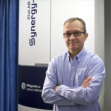
The third episode of the webinar series on electron diffraction offers a comprehensive, practical guide to the entire microED (3DED) workflow for structural researchers, emphasizing its accessibility and power in solving submicron crystal structures. Presented by Dr. Jessica Burch, the session details each phase of an electron diffraction experiment using the XtaLAB Synergy-ED diffractometer, focusing on powders that are not traditionally analyzed by single crystal X-ray diffraction (SCXRD).
Jessica opens with an overview of 3DED, highlighting how dynamical scattering is not as prohibitive as traditionally expected, especially when performing continuous rotation electron diffraction. This shift enables kinematical structure solutions—analogous to SCXRD methods—despite the strong interaction between electrons and matter. The practical advantage is that researchers can now analyze individual grains within a polycrystalline powder, potentially resolving multiple components, polymorphs, or phases, even from nanogram-scale or air-sensitive samples.
The talk emphasizes the use of standard transmission electron microscopy (TEM) grids and intuitive sample prep methods. Sample requirements include crystallinity and submicron thickness. Powder is deposited onto the grid through simple techniques like gentle pressing and shaking, and Jessica provides guidance for optical pre-characterization and size estimation. She also shows how cryogenic handling mitigates vacuum-induced degradation, crucial for solvent-labile or metastable phases.
Jessica details the XtaLAB Synergy-ED’s integrated workflow, which simplifies traditionally complex TEM alignment procedures and separates it from the demands of conventional EM usage. Visual and diffraction modes allow for real-time screening of particles, with immediate feedback on unit cell parameters and quality. The software suite, CrysalisPro coupled with AutoChem and Olex2, allows seamless progression from beam alignment and data collection to structure solution and refinement.
She also introduces automation tools such as queuing and results viewer modules, enabling unattended high-throughput data collection and facilitating grain-by-grain screening of complex powder mixtures. Automation not only improves efficiency but supports statistical exploration of mixtures, including clustering based on unit cell parameters and automated merging of data sets from low-symmetry crystals.
Throughout, Jessica underscores the growing maturity of electron diffraction as a mainstream structural method. The system democratizes access to advanced crystallography for non-specialists, moving ED from a niche, expert-driven method to a routine, rapid, and high-fidelity solution for diverse structure determination challenges.
Key questions answered in the webinar
-
Electron diffraction is a technique that uses electrons as the radiation source to analyze the structure of crystalline materials. It complements X-ray diffraction by allowing researchers to study crystals that are too small to analyze with single crystal X-ray diffraction, particularly those smaller than one micron. Electrons interact much more strongly with matter than X-rays, making them suitable for studying these submicron-sized crystals.
-
Traditionally, electron diffraction involved aligning a crystal to a zone axis and recording a single diffraction pattern, which maximizes dynamical scattering and makes structural solution difficult. The newer 3D ED or micro ED approach, developed around 2007, involves continuously rotating the crystal during data collection, similar to single crystal X-ray diffraction. This continuous rotation minimizes dynamical scattering, allowing the use of the kinematical approximation for structural solution and refinement, making it more akin to X-ray crystallography workflows.
-
Samples suitable for 3D ED/micro ED must be crystalline and have particle thicknesses of less than one micron. While traditional X-ray diffraction is often preferred for larger crystals, ED is particularly useful for analyzing powdered materials, allowing for grain-by-grain analysis of mixtures, impure samples, air-sensitive materials, and samples available in only nanogram amounts or requiring solvation. A powder X-ray diffraction pattern with peaks past 30° 2θ (using copper radiation) is often a good indicator of crystallinity suitable for ED.
-
Sample preparation for ED typically involves placing a fine powder of the material onto a commercially available TEM grid (usually 3 mm in size) with a metal mesh and an electron-transparent film (like continuous carbon or lacy carbon). A common method is to gently tap the grid into the powder to transfer grains, then shake off the excess. The grid is then secured onto a sample holder and inserted into the diffractometer's vacuum chamber. For vacuum-sensitive samples, cryogenic sample loading using a cryo-holder precooled with liquid nitrogen is employed to prevent loss of solvent.
-
For successful data collection, crystallites on the TEM grid should be less than one micron in thickness. It's also beneficial to have crystallites oriented in various directions (avoiding preferred orientation) to maximize the completeness of the collected data within the goniometer's rotation range (typically around 160°). Additionally, crystallites should be spaced a few microns apart to allow for rotation without being obstructed by other crystals or grid bars.
-
The manual data collection workflow starts with locating suitable crystals in visual mode using a low magnification overview (mini map) and then zooming in. The user then switches to diffraction mode to assess the quality of the diffraction pattern from selected crystallites. If a crystal diffracts well, the user centers it, determines the maximum tilt range free from obstructions, sets up the rotation experiment parameters (exposure time, tilt range), and initiates the data collection. The system then collects a series of diffraction images as the crystal rotates.
-
Automation significantly streamlines the ED workflow through features like queuing, which allows for unattended data collection on multiple user-identified particles. This involves setting up pre-experiments (quick snapshots to assess diffraction quality) and full experiments for data collection. The system can automate tasks like centering the crystal within the aperture and determining the maximum unhindered tilt range through eccentric height adjustment and occlusion detection. Automated processing software (like AutoChem) runs concurrently with data collection, providing real-time feedback on the unit cell and potential structural solutions, allowing users to quickly evaluate data quality and decide whether to continue collecting or move to another grain.
-
After data collection, the raw diffraction images are processed (indexed, reduced, and potentially solved) using integrated software like AutoChem and refined in programs like Olex2. The results are then viewed and managed using a results viewer, which helps organize potentially large volumes of data from multiple experiments. Merging multiple datasets is frequently necessary in electron diffraction, especially when crystals exhibit preferred orientation, to achieve a complete dataset covering all necessary reflections for a robust structural solution. Tools are available to aid in selecting datasets for merging and automating the merging process.

Contact Us
Whether you're interested in getting a quote, want a demo, need technical support, or simply have a question, we're here to help.
