X-ray Microscopy Seminar and Workshop
Wednesday, March 30, 2022 @ University of Delaware
In this seminar, we invited top-level innovators and researchers in the field to learn the latest advances in X-ray microscopy, including an application of deep learning to X-ray CT image analysis and how you can apply X-ray CT to various research areas.

- Program
- Presenters
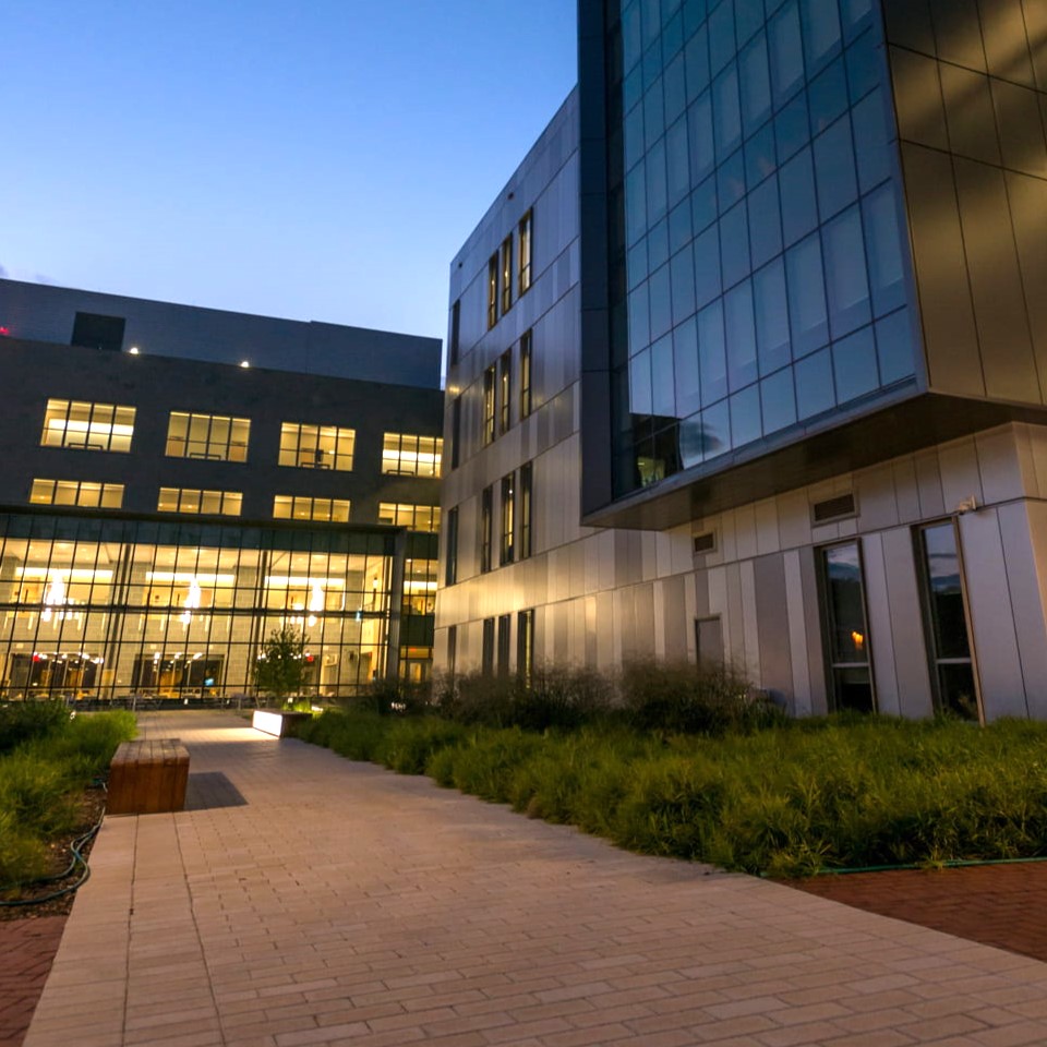
- Location
- Recording
Seminar and workshop program

Collaborators: Shivam Chauhan, Erik Ervin, and Paul Imhoff

Abstracts
Opening Remarks - Recording
Dr. Anshuman Razdan (Associate Vice President, Research and Development at the University of Delaware)
Micro-CT Scanning with 3D Image Analysis for Porosity Studies of Historic Bricks and Archaeological Ceramic Sherds - Recording
Chandra L. Reedy (Invited speaker)
Professor and Director,
Center for Historic Architecture and Design
University of Delaware
Characterizing pore systems in historic bricks and archaeological ceramics reveals information about the selection and processing of raw materials, production and firing technologies, functional use properties, degree of deterioration, and effectiveness of preservation measures and conservation treatments. Some of the data of interest includes total porosity, ratio of pores accessible to the surface versus inaccessible interior ones, range of pore sizes and shapes, various pore connectivity measures, and the spatial relationship of pores to particle inclusions. 3D image analysis (using the Dragonfly software package) of micro-CT scans of historic American bricks and ancient Chinese ceramic sherds will be discussed. Some of the issues that need to be considered include sample size and spatial resolution versus representativeness, variability and replication, and choice of variables to measure. The deep learning segmentation approach discussed includes intensity calibration of images and the use of comparative images to aid segmentation decisions (2D petrographic thin sections, and micro-CT images improved through a combination of image filters or by Noise2Noise denoising). Some of the imaging challenges posed by these objects will be highlighted.
Microstructural Analysis of Carbon-Carbon Composites During High-Temperature Processing - Recording
Faheem Muhammed (Student speaker)
University of Delaware
As a result of the time-intensive re-densification process, there is a large cost associated with the fabrication of carbon-carbon composites. It is hypothesized that controlling the composite's microstructure during the carbonization stage may lead to an increased permeability during re-impregnation, which would reduce the number of steps required for full consolidation, and lead to an overall reduction to the volume of inaccessible voids in the final composite. To begin to develop a framework to determine the most desired void microstructure to maximize the efficiency of re-infiltration, a kinetics driven approach was utilized to determine the various stages of void and porous network formation. Through thermogravimetric analysis, and subsequent kinetic modeling, it was possible to relate the progression of pore formation (void growth, the progression of crack density, and three-dimensional pore network formation) to the extent of thermal decomposition. To understand the progression of the pore morphology, X-Ray Computed Tomography was utilized in order to perform quantitative analysis, as a function of the degree of conversion of the matrix material. By utilizing the built-in functionality of Dragonfly, information regarding the average void diameter, pore interconnectivity, and the cessation of void growth was possible.
Deep Learning Automated Image Segmentation - No expertise required - Recording
Mike Marsh (Invited speaker)
Dragonfly Product Manager
Object Research Systems
We will look at examples of how a user-friendly Deep Learning platform can fully automate image segmentation on images that would otherwise take countless hours of effort to fully label. We'll see how a variety of biological and materials science samples are easily managed by Deep Learning, even by users who have no expertise or background in machine learning/deep learning. Finally, we'll explore some questions and best practices in how to optimize performance so users can get highly robust models with minimal effort spent preparing ground truth.
Understanding the Impact of Biochar Amendment on Macropores in Soil from 3D X-Ray CT Tomography - Recording
Marcus Bowser* (Student speaker), Shivam Chauhan**, Erik Ervin***, and Paul Imhoff*
* Department of Civil and Environmental Engineering
University of Delaware, ** 2Department of Chemical and Biomolecular Engineering, University of Delaware *** 3Department of Plant and Soil Sciences, University of Delaware
Urban soils are typically highly compacted, so when a storm event occurs and stormwater drains onto these soils from impervious surfaces, e.g., roofs and roads, the water cannot infiltrate well into the ground and instead drains directly into the stormwater treatment system. This stormwater runoff causes increased loading of pollutants and water, requiring significant investment in stormwater treatment infrastructure to achieve the increasingly stringent EPA regulations. Biochar has been proposed as a soil amendment to improve water retention, stormwater infiltration, and plant growth in urban soils to mitigate this issue. Biochar amendment usually increases soil aggregate size and porosity through biological processes that are not entirely understood, which were correlated with 50 to 500% increases in stormwater infiltration in recent field trials. Unfortunately, the linkage between biochar-induced increases in soil aggregation and porosity and pore network changes that improve water flow has not been established. We are using high-resolution X-ray CT and 3D image analysis to evaluate the impact of biochar amendment on pore networks in soils with varying amounts of biochar, under low and high compaction, and with or without turf grass as soils age. The soil pore networks deduced from X-ray CT and 3D image analysis allow us to understand the dynamic changes in pore networks in biochar amended soils and to link this change to smaller-scale changes in soil aggregation.
Modeling Sustained Release through Non-destructive Computed Tomography - Recording
Xutao Shi (Student speaker)
University of Delaware
Sustained-release systems improve the bioavailability of biopharmaceutical products and eliminate the necessity for a frequent administration schedule of the drug. A better understanding of such delivery systems, and therefore a more efficient system design, can be achieved by using mathematical models capable of predicting the drug release profiles a priori. However, these models are often formulated phenomenologically, and morphological details such as the early-stage pore formation of the sustained-release system are often neglected or assumed without proper experimental characterization. In this work, we utilized non-destructive X-ray computed tomography (CT) to quantitatively characterize the internal structure of a PLGA-based sustained release system. We modified an existing mechanistic sustained-release model and incorporated the structural characteristics obtained from segmentation of the three-dimensional CT reconstructions. The model predictions were compared to the release profiles obtained from experimental studies. The results demonstrate the importance of structural porosity in sustained release and the potential of a CT-aided mechanistic model to predict the drug release profiles of a PLGA-based sustained-release system.
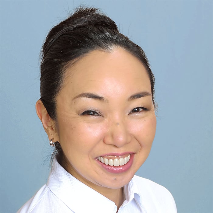
Basics of X-ray Microscopy - Recording
Dr. Aya Takase
Head of Global Marketing Communications
Rigaku
This talk gives you an overview of X-ray computed tomography, including the basics of X-ray imaging, instrumentation, data analysis, and application examples.
Questions?
Contact us at info@rigaku.com.
Contact Us
Whether you're interested in getting a quote, want a demo, need technical support, or simply have a question, we're here to help.

Subscribe to the X-ray CT Email Updates newsletter
Stay up to date with CT news and upcoming events and never miss an opportunity to learn new analysis techniques and improve your skills.
