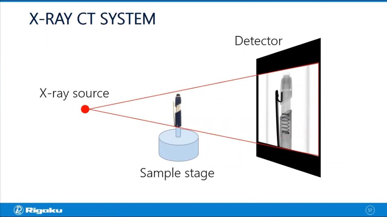
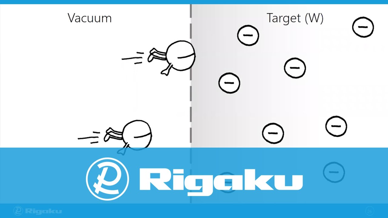
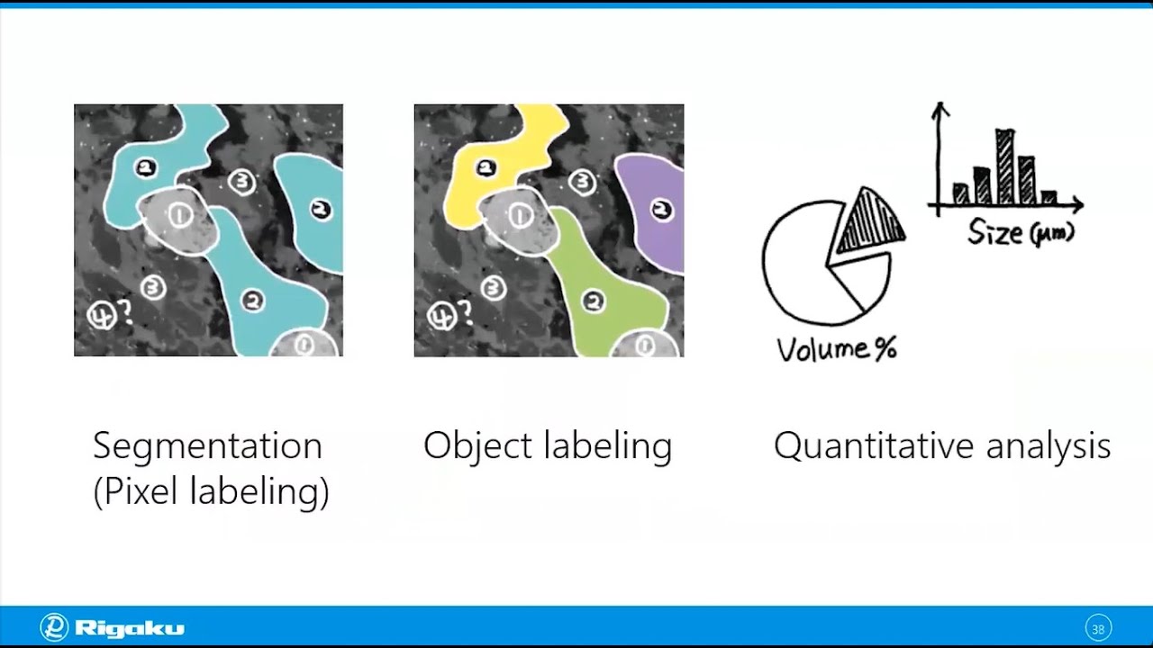
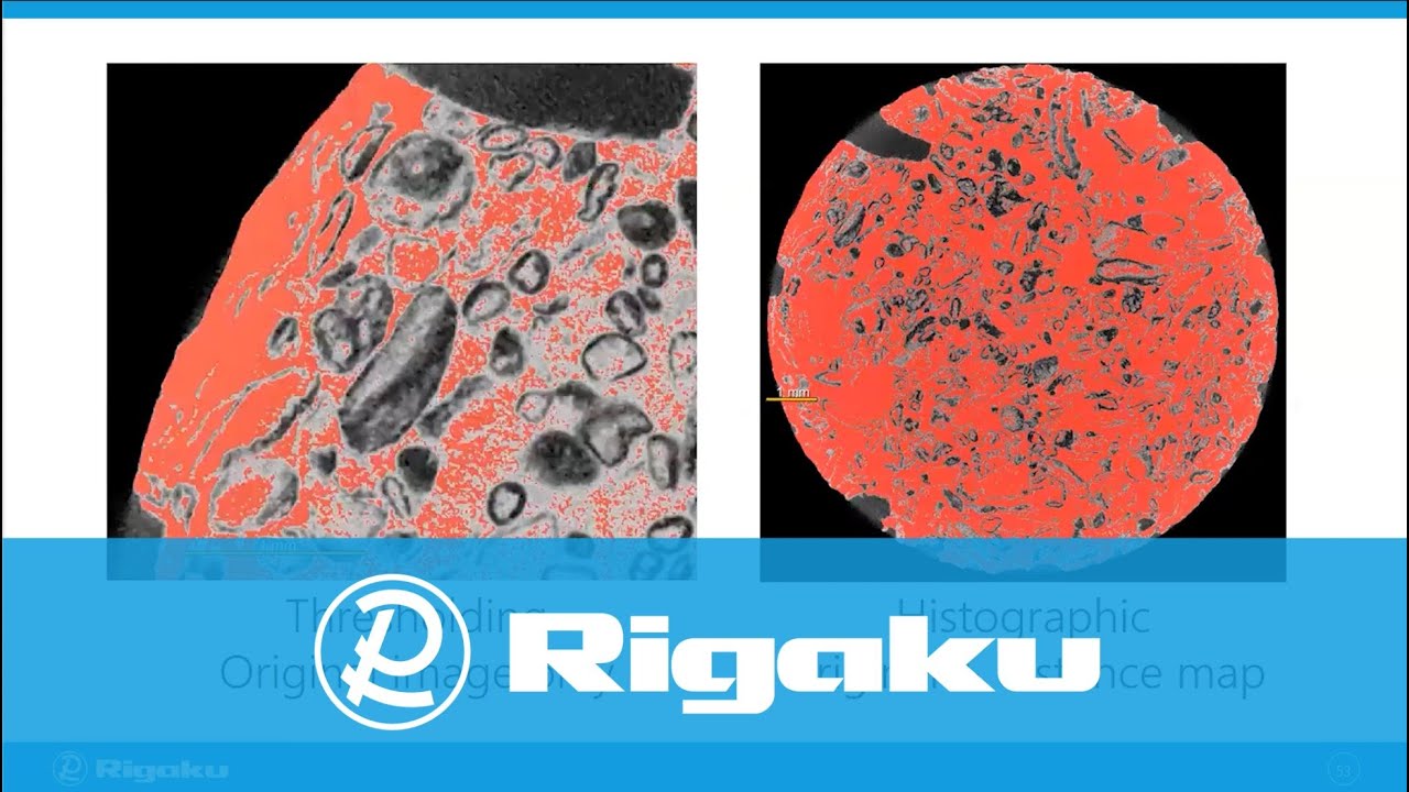
X-ray CT Menu

X-ray Computed Tomography for Materials & Life Sciences

Learn how X-ray computed tomography (CT) works, how the results are analyzed, and how this X-ray imaging technique is applied to materials and life science research.

Introduction

This is a good place to start if you are new to X-ray computed tomography (CT). In this webinar, you will learn how X-ray CT works and how it can be applied to scientific research. It will introduce the technique, instrumentation, and application examples.

Rigaku
Head of Global Marketing Communications

Data Analysis

There are many ways to analyze CT images, and the process of analyzing images and extracting quantitative information can be involved. This webinar will provide an overview of CT data analysis, including thresholding segmentationmachine learning-based segmentation, and quantitative analysis. Several commonly used data analysis and visualization programs will be discussed and demonstrated to help you get an idea of where to start and select the right analysis tool for your needs.

Rigaku
Head of Global Marketing Communications

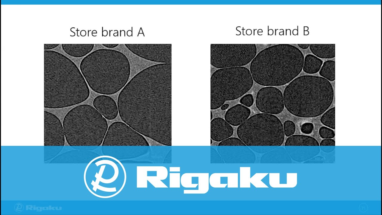
Food and Pharmaceutical Applications

You will see many X-ray CT application examples in the food and pharmaceutical industries in this webinar, including the analyses of cracks and aggregation inside tablets, tablet and drug particle coating thicknesses, air pocket size distributions in food, and sugar coating thicknesses of candies.

Rigaku
Head of Global Marketing Communications

Foams and Composites Applications

You will see many X-ray CT application examples of foams and composite materials in this webinar, including the analysis of foam porosity, cell size, and cell wall thicknesses as well as void distribution and fiber orientation in composite materials.

Rigaku
Head of Global Marketing Communications

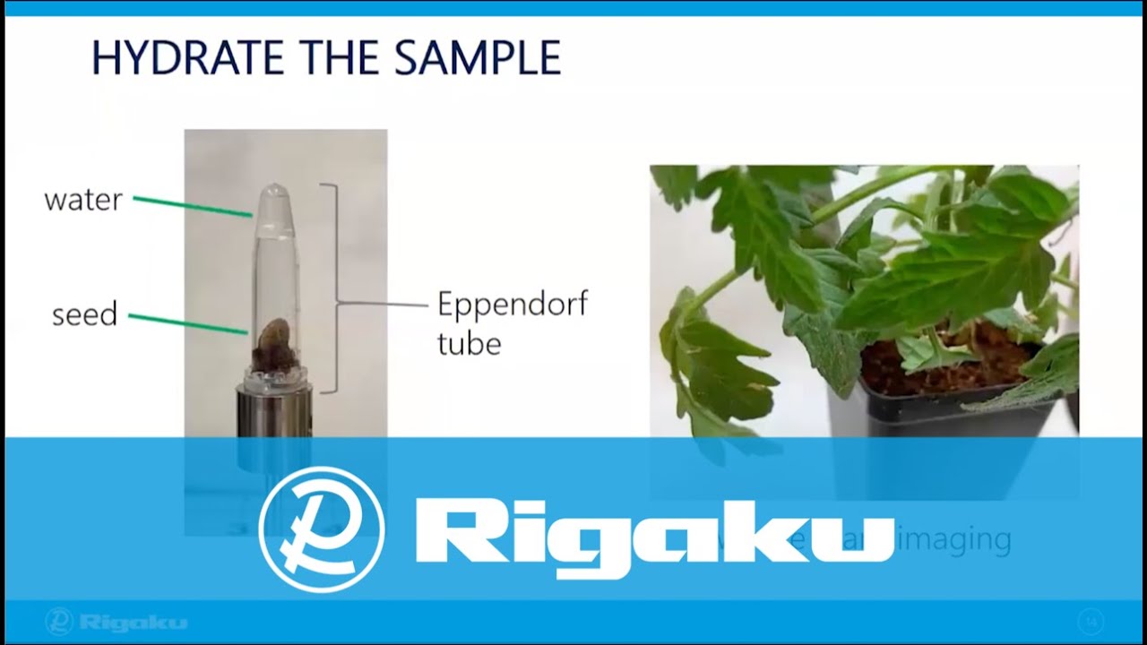
Plant Science Applications

You will see many examples of X-ray CT application for plants and seeds in this webinar, including the non-destructive characterization of plant traits for ripening fruit, seeds, and root systems. Additionally, an analysis of cell wall thicknesses and void distribution among different seed varieties is presented.

Rigaku
Director of X-ray Imaging

Geology Applications

X-ray CT has become an essential tool for geological sample analysis. You will see many application examples in this webinar, including the analysis of cracks, pores, inclusions, and phase quantification of rocks and drill cores. We also introduce available resources for pore network analysis that can be applied to rock CT scans.

Rigaku
Head of Global Marketing Communications

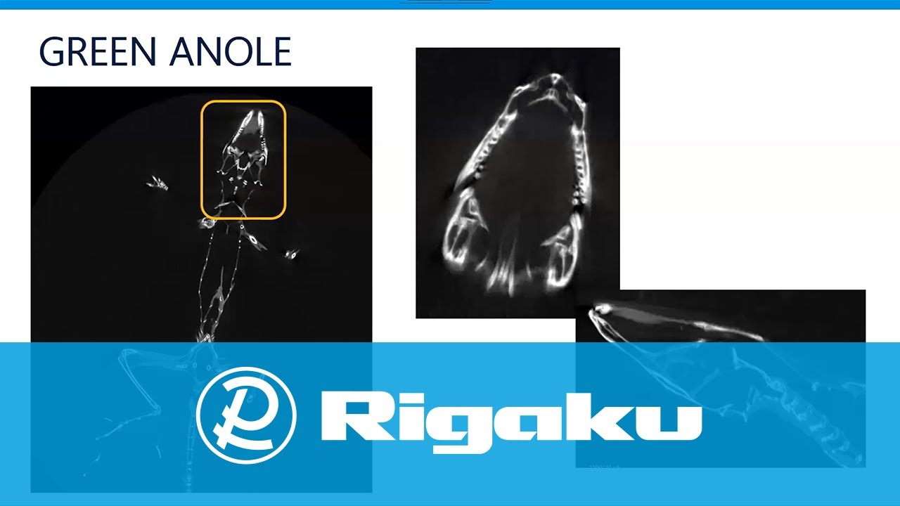
Life Science Applications

In this webinar, you will see 3D views of the structures of a reptile, insects, and a mouse, including their stained organs. You will also learn how to deal with unique challenges in life science sample preparation and how to conduct quantitative analyses.

Rigaku
Director of X-ray Imaging

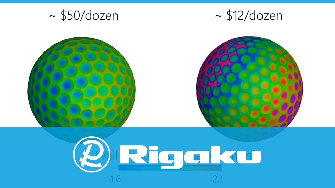
Metrology Applications

This webinar presents the basics of metrology analysis and a number of X-ray CT application examples. Examples include size and shape measurements of metal and plastic parts, tolerancing evaluation, comparison of nominal (CAD) and actual (CT) or a golden standard, and a test subject. We also introduce available resources to learn more about X-ray CT metrology. 

Rigaku
Head of Global Marketing Communications

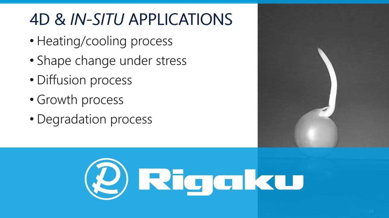
4D and In-situ Applications

In this webinar, you will learn the keys to successful 4D and in-situ X-ray CT measurements and how to plan experiments. You will also see many examples of applications, including the observation of foam under compression, the drug degradation process, and the seed germination process. 

Rigaku
Head of Global Marketing Communications

Preparing Life Science Samples

In this webinar, you will learn sample preparation techniques for life science samples. These samples typically require preservation to ensure that ‘as lifelike as possible’ CT data can be collected. These samples typically have low-density contrast and thus benefit from staining. You will see different preparation techniques and application examples.

Rigaku
Director of X-ray Imaging

Contact Us

Whether you're interested in getting a quote, want a demo, need technical support, or simply have a question, we're here to help.

Subscribe to the X-ray CT Email Updates newsletter

Stay up to date with CT news and upcoming events and never miss an opportunity to learn new analysis techniques and improve your skills.