Application Note PX015
Introduction
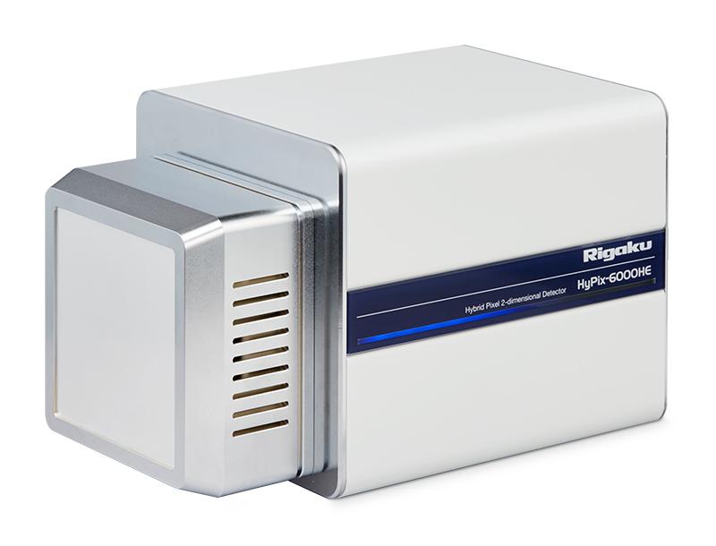
The HyPix-6000HE hybrid photon counting (HPC) detector (Figure 1) is the latest detector for protein data collection designed by Rigaku. The HyPix-6000HE detector possesses all the benefits associated with HPC detectors: high sensitivity, large dynamic range, very low read noise, single pixel point-spread function and shutterless data collection.

Figure 1: The HyPix-6000HE HPC detector.
In addition, the HyPix-6000HE also features a small pixel size of 100 μm x 100 μm and a frame rate of 100 Hz. The small pixel size of the HyPix-6000HE facilitates improved spatial resolution for reflections from crystals with long unit cell axes, and enables data collection with shorter detector distances. The high frame rate of 100 Hz allows a coincidence correction for high count rates and greatly improves the accuracy with which reflection intensities are measured, especially for the strong reflections. Lastly, the HyPix-6000HE is air-cooled, thereby lowering its maintenance costs. The combined features of the HyPix-6000HE allow fast and high accuracy data collection for single crystal samples
Spatial resolution for thaumatin reflections
Data were collected on crystals of the standard protein thaumatin from Thaumatococcus danielli (Figure 2). This protein was selected because crystals are robust and the protein crystallizes easily in a tetragonal space group with unit cell parameters 58 x 58 x 150 Å.

Figure 2: Thaumatin (PDB ID: 1RQW).¹
One experimental goal was to explore the detector distance at which one can reasonably collect data where reflections along the longest unit cell axis (150 Å) are spatially resolved on the detector. A general recommendation for data collection on HPC or CCD detectors is to collect, in mm, at one half the longest primitive unit cell distance, in Å, which would be approximately 75 mm for the case of thaumatin. To test, data were collected from a small thaumatin crystal (~50 x 50 x 80 μm) using 30 sec/0.3° exposure times at three different detector distances: 80 mm, 60 mm and 41 mm. The data were collected on a Rigaku MicroMaxTM-007 HF microfocus rotating anode, using Cu Kα radiation, configured with a kappa goniometer and the HyPix-6000HE HPC detector.
The diffraction image obtained at a distance of 80 mm is shown in Figure 3a. The
red rectangle outlines a row of reflections along the longest unit cell edge of 150 Å. This area is zoomed in for easier display in the right hand column for all three detector distances: 80 mm (3b), 60 mm (3c) and 41 mm (3d). The zoomed pictures show that, while the number of background pixels between reflections decreases with detector distance, reflections are still resolvable on the detector with one or two background pixels between reflections. This is true even at 41 mm. A 2D intensity profile plot (3e) is shown for the depicted reflections collected at 41 mm, showing that the intensity returns to background levels between adjacent reflections, further confirming that the reflections are resolved.

Figure 3: a): Diffraction image for thaumatin collected at 80 mm. Zoomed display of a row of reflections along the longest unit cell edge of 150 Å at b) 80 mm, c) 60 mm and d) 41 mm. e) 2D profile for reflections shown in ‘d)’. Text in blue depicts the distance at which the data image was collected.
Structure solution of thaumatin by molecular replacement
Data were collected for a larger thaumatin crystal (~150 μm x 100 μm x 100 μm), using CrysAlisPro, at a crystal-to-detector distance of 80 mm (Table 1).² Figure 4 shows the first image of the data set.

Figure 4: First diffraction image of the data set collected on thaumatin on the HyPix-6000HE.
Data were processed with XDS and data statistics are shown in Table 2.³
Table 1: Data collection parameters used to collect thaumatin data on the HyPix-6000HE detector. The total range of data collected was 135 with a total X-ray dose time of 2 hours and 30 minutes.
| Scan ID | Exp | Δω | Dist | 2θ | φ | κ | # img |
| 1 | 10 sec | 0.15º | 80 mm | -18.54° | 0º | 178º | 400 |
| 2 | 10 sec | 0.15º | 80 mm | -18.54° | -60º | -57º | 500 |
Table 2: XDS processing statistics.
| Parameter | Value |
| Space Group | P4₁2₁2 |
| Resolution cutoff (Å) | 2.05 |
| Completeness (last shell) (%) | 99.0 (98.0) |
| Redundancy (last shell) | 3.9 (3.0) |
| <I>/<σ(I)> (last shell) | 16.7 (2.3) |
| CC (1/2) | 99.7 (76.3) |
| Rfac (last shell) (%) Rmeas (last shell) (%) |
6.5 (47.6) 7.5 (58.0) |

Figure 5: 2Fo-Fc electron density map contoured at 1.5σ.
The crystal structure of thaumatin was solved by molecular replacement in space group P4₁2₁2 using HKL-3000R®.⁴ HKL-3000R used MOLREP for the rotation and translation functions search, ARP/wARP to trace the protein and place the waters, and REFMAC to refine the model.⁵,⁶,⁷ MOLREP was run with data to 3.0 Å, using PDB ID ‘1RQW’ as the starting model, from which the side chains, ligands and waters had been removed. The model was then subjected to 5 cycles of rigid body refinement with Refmac using data to 3.0 Å, followed by 10 cycles of restrained atomic refinement using the entire data set to 2.05 Å. Using the sequence from PDB ID ‘1RQW’, 97% of the model (201 residues out of 207) was built after 3 auto-building cycles in ARP/wARP for Rfac and Rfree values of 20.2% and 26.5%, respectively.
The model was then submitted to 10 additional refinements in Refmac, followed by manual adjustment and addition of a few side chains into their electron density. Final refinements yielded Rfac and Rfree of 18.6% and 24.6%, respectively, for a Figure of Merit of 85.4%. A representative area of the final 2Fo-Fc electron density map is displayed in Figure 5.
Conclusion
The HyPix-6000HE HPC detector from Rigaku was tested with thaumatin using the MicroMax-007 HF microfocus rotating anode. These results show that the combination of 100 μm pixel size with the single pixel point spread function of the HyPix-6000HE allows one to resolve reflections along the long unit cell edge of thaumatin (150 Å) at a detector distance of 41 mm. Additionally, the HyPix-6000HE is shown to collect high quality data for thaumatin, which allowed for structure solution and refinement of the crystal structure of thaumatin down to Rfac and Rfree of 18.6% and 24.6%, respectively. These results illustrate the effectiveness of the HyPix-6000HE detector to resolve reflections along longer unit cell edges and accurately collect data for structure solution and refinement. These qualities, combined to the usual features of HPC detectors and easy maintenance, makes the HyPix-6000HE a great detector to quickly screen and collect data on a wide variety of samples in the home lab.
References
- Ma Q., Sheldrick G. M. (2003) doi: 10.2210/pdb1rqw/pdb
- Rigaku Oxford Diffraction, (2016), CrysAlisPro Software system, version 1.171.39.7d, Rigaku Corporation, Oxford, UK.
- Kabsch W. (2010a) Acta Cryst. D66, 125-132.
- Minor W., Cymborovski M., Otwinowski Z., & Chruszcz M. (2006) Acta Cryst. D62, 859-866.
- Vagin A.A. & Teplyakov A. (1997) J. Appl. Cryst. 30, 1022-1025.
- Langer G., Cohen S.X., Lamzin V.S., Perrakis A. (2008) Nat Protoc. 3 (7):1171-9.
- Murshudov G.N., Vagin A.A., & Dodson E.J. (1997) Acta Cryst. D53, 240-255.

