Application Note SMX037
Introduction
Single-crystal electron diffraction (SC-ED), also known as microED/3DED offers access to smaller samples than are possible with X-ray diffraction techniques. Whilst currently available home lab X-ray sources offer the possibility to study samples down to only 1 micron in their smallest dimension¹, in many cases it is difficult or impossible to grow crystalline material of sufficient volume and quality to meet this requirement. Cases where material is limited or of lower quality are particularly challenging.
One such field where crystallisation can be an obstacle is determining the stereochemistry of degradation products of biologically active compounds such as fungicides.
Electron diffraction can provide three-dimensional structural information on samples as small as a few tens or hundreds of nanometres in thickness. This makes it a valuable tool for cases where the above issues exist.
The Syngenta product SOLATENOL™ (Figure 1) is a commercial fungicide used in agriculture. A degradant of this compound was selected in order to demonstrate the ability to resolve stereochemistry by diffraction methods. This sample was analysed after synthesis and purification, with no attempts at crystallisation, using only a few milligrams of sample.
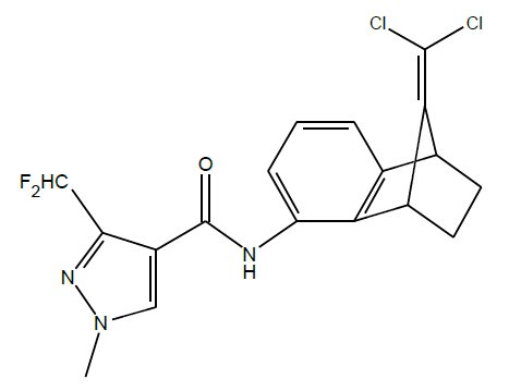
Figure 1: The Syngenta fungicide, SOLATENOL
Instrumentation
The XtaLAB Synergy-ED from Rigaku (Figure 2) is a single crystal electron diffractometer that combines core technologies from Rigaku Corporation and JEOL Ltd. The XtaLAB Synergy-ED is designed to be easy to use by X-ray crystallographers and newcomers to electron diffraction alike. Our HyPix-ED detector (Figure 3) offers high-sensitivity and low noise characteristics to enable high quality measurement of data from extremely small samples.
.jpg?width=800&height=610&name=XtaLAB%20Synergy-ED%20electron%20diffractometer%20(doors%20closed).jpg)
Figure 2: The XtaLAB Synergy-ED.
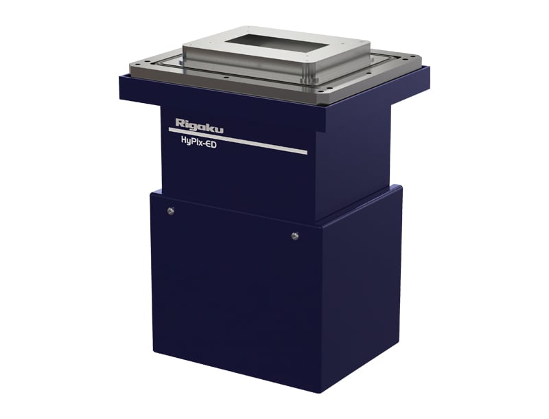
Figure 3: The HyPix-ED, an HPAD detector with high sensitivity and low noise for electron diffraction experiments.
The XtaLAB Synergy-ED is controlled by CrysAlisPro, enabling many of the features and functionality designed for X-ray diffraction to be directly applied to electron diffraction, such as AutoChem (Figure 4), which enables concurrent automatic structure solution and refinement while the experiment is still in progress. The XtaLAB Synergy-ED therefore offers an intuitive product, which is easy to integrate into existing analytical services departments.
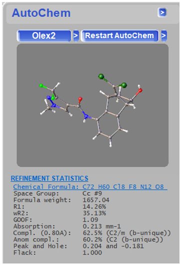
Figure 4: AutoChem allows structures to be obtained automatically for in-progress data collections.
Experimental
Data collection on the XtaLAB Synergy-ED is straightforward. From this sample, two datasets were collected on two different grains and then merged to produce a higher completeness, composite dataset. Data were collected at room temperature from grains approximately 100 nm in thickness. Images of the grains can be seen in Figure 5.
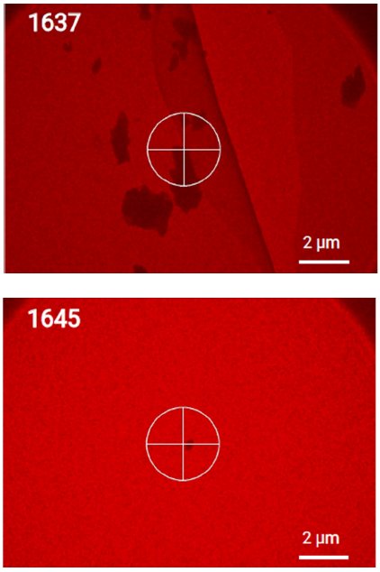
Figure 5: Samples chosen for the SC-ED experiments shown prior to centring to better indicate approximate size.
Electron diffraction experiments are typically very fast, consisting of a single axis scan up to approximately 160° depending on the sample environment. Total data collection time was only 4 minutes using 0.5 s exposure times and 0.5° frame width as shown in Table 1.
Table 1: Data Collection details.
| Data | Number of frames | Scan width (°) | Exposure time (s) |
Total time |
| 1637 | 200 | 0.5 | 0.05 | 00:01:50 |
| 1645 | 240 | 0.5 | 0.5 | 00:02:10 |
| all | 440 | - | - | 00:04:00 |
Data were processed using CrysAlisPro and structures were refined using kinematical methods and SFAC instructions suitable for electron diffraction. The composite dataset provided good statistics and reduced the influence of dynamical scattering effects. Data statistics are summarised in Table 2.
Table 2: Selected data statistics.
| Data | 6877 |
| Completeness (%) | 90.2 |
| Redundancy | 4.4 |
| <F²> | 737.77 |
| <F²/σ(F²)> | 11.62 |
| Rpim (%) | 12.9 |
| Resolution limit (Å) | 0.83 |
Note that from only 4 minutes of data collection time on samples of only a few hundred nanometres thickness, > 90 % completeness was achieved to a resolution of 0.83 Å.
All non-hydrogen atoms were refined anisotropically. The structure obtained is shown in Figure 6.
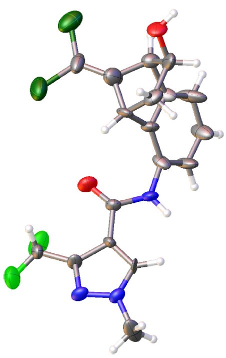
Figure 6: The structure of the SOLATENOL™ degradant showing relative stereochemistry of the OH group.
Discussion
In order to bring biologically active products such as fungicides to market, metabolites and other degradants must be fully characterised along with the active compound so that hazards can be predicted or identified and risk-assessed. Since proteins are chiral, the stereochemistry of the active compound and its metabolites plays a key role in activity and as such, understanding this as well as the connectivity is very important. For the molecule considered here, stereochemistry had been previously determined through a combination of techniques, which was time-consuming. A crystal structure however, would have addressed most of the characterisation needs in a single experiment, with a high degree of certainty.
Conclusions
With the newly available XtaLAB Synergy-ED, single-crystal electron diffraction experiments were possible on this SOLATENOL™ degradant.
In this case, the XtaLAB Synergy-ED provided data and a crystal structure that was simply not possible using conventional single-crystal X-ray diffraction due to the sample size. Data collection time was only a few minutes and the critical information (the stereochemistry) was clearly revealed as shown in Figure 6.
Authors
- Peter Howe, Mark Montgomery and Jenny Moore, Syngenta AG
- Fraser White, Rigaku Oxford Diffraction

