High-speed RSM of a III-nitride Epitaxial Film by 1D Detection Mode
Introduction
Reciprocal space mapping (RSM) is an XRD technique used to evaluate lattice spacing and crystal orientation distribution independently from each other, applied to the analysis of thin film samples such as epitaxial films. Since a reciprocal space map requires multiple scans with various combinations of the scattering angle (2θ) and the incident angle with respect to the sample (ω), it can take a relatively long time to collect the necessary data in general. Combination of the 1D exposure mode of a 2D detector and high-speed scanning by the ω axis enables data collection in a very short time, from several tens of seconds to several minutes.
Measurement and analysis
Reciprocal space mapping was performed for a thin film sample, a [GaN (15.2 nm) / In₀.₀₈Ga₀.₉₂N (3.5 nm)] x5 multiple quantum well (MQW) on a sapphire substrate. Figure 1 shows the observed data from a high-speed ω scan using a rectangular 2D detector, installed vertically at a detector distance of 300 mm. The scan duration was 30 seconds. Since the detector operated in 1D exposure mode, it could capture diffraction signals over a wide 2θ range (calculated from its sample to detector distance and the length of its longitudinal detection) without moving the 2θ axis. An ω scan alone collected enough data to construct the reciprocal space map.
Figure 2 shows the overlay view of the reciprocal space map generated by converting the data shown in Figure 1 into reciprocal space coordinates and simulated reciprocal lattice points based on the structure model of the sample. It was confirmed that the observed reciprocal lattice points matched the 0th-order reflections of the sapphire substrate and InGaN, and satellite peaks due to the MQW aligned vertically.
As described above, this technique enables the measurement of a reciprocal space map in a range sufficient to observe reciprocal lattice points of the substrate and the epitaxial layers of a typical epitaxial film sample within only several tens of seconds to several minutes.
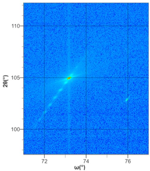
Figure 1: High-speed ω scan data (1D exposure mode)
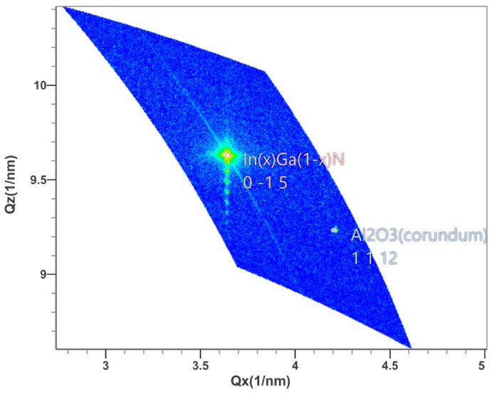
Figure 2: Observed reciprocal space map and simulation
Reference
T. Kuzumaki and A. Ogi: Rigaku Journal (English version), 34 (2) (2018) 17-20.

Contact Us
Whether you're interested in getting a quote, want a demo, need technical support, or simply have a question, we're here to help.