Visualization of Components in Ceramic Composites using a High-resolution 3D X-ray Microscope
Introduction
Composite materials are made by combining two or more different materials. X-ray computed tomography (CT) visualizes differences in X-ray absorbance in an object as contrast in the image. For example, ceramics, resin, and voids are represented by different gray levels. Rigaku’s high-resolution 3D X-ray microscope, nano3DX, an X-ray CT instrument, is capable of acquiring images of a sample at sub-micron-scale resolution and showing a zoomed image of the sample’s internal structure in three dimensions. This allows you to understand the dispersion of each component that affects the properties of the composite, presence/absence of separation at the boundary surface of the material, and the size of the voids. In this example, the components of a ceramic composite were visualized and the volume ratio was calculated.
Measurements and results
A ceramic composite was scanned for 2 hours using a high-resolution 3D X-ray microscope with a Mo source. The cross-section image of the CT reconstruction results represents SiO₂ particles, CaCO₃ particles, resin, and voids contained in the ceramic composite with different gray levels (Figure 1). The CT image was segmented based on the gray levels and colored by component. Furthermore, the volume fractions of each component and voids were calculated from the sum of the volume of the three-dimensional pixels (Figure 2). In this way, the high-resolution 3D X-ray microscope enables the visualization of a few to several tens of micrometer-scale particles contained in a composite material in three dimensions.
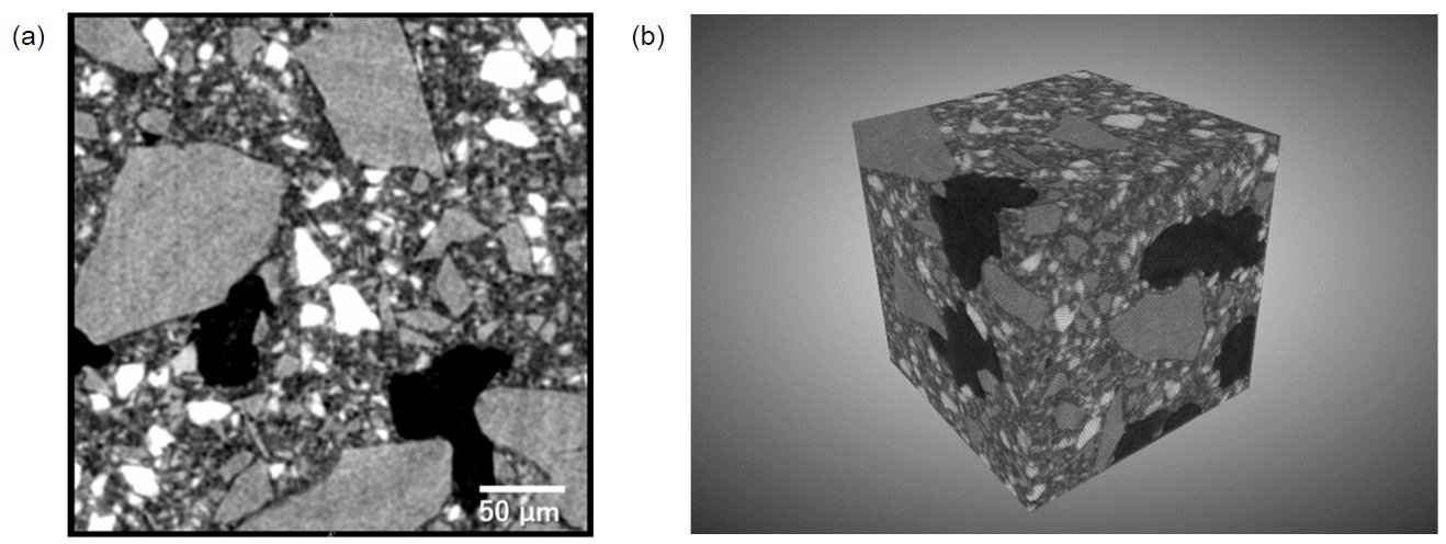
Figure 1: CT reconstruction results of a ceramic composite: (a) CT cross-section image, (b) 3D image (300 μm on a side)
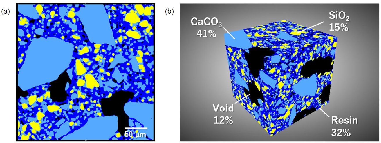
Figure 2: Segmentation results of ceramic composite: (a) CT cross-section image (b) 3D image and volume fraction of each component (300 μm on one side)

Contact Us
Whether you're interested in getting a quote, want a demo, need technical support, or simply have a question, we're here to help.