CT Observation of a Laminated Battery Cell by X-ray Microscopy
Introduction
Batteries are made of diverse materials. Non-uniformity, impurities, and defects within the battery structure negatively affect the performance, stability, and durability of the product. As batteries are repeatedly charged and discharged, their quality degrades due to changes in the internal structure. Therefore, to evaluate a battery’s internal structure, a non-destructive observation technique is needed. By using an X-ray microscope, which can observe transparently the internal area of a sample, the micron structure within a laminated battery cell can be observed in a non-destructive manner. The structure can be visualized in 3D as well by using the CT (Computed Tomography) technique.
Measurements and results
A CT measurement was performed using the nano3DX X-ray microscope on a 10 mm wide single-layer lithium ion battery cell laminated with aluminum (Figures 1 and 2). As a result, the active materials and carbon particles within the battery cell were clearly rendered. Structural changes during charging and discharging can also be observed in a non-destructive manner by attaching an electrode to the laminated cell.
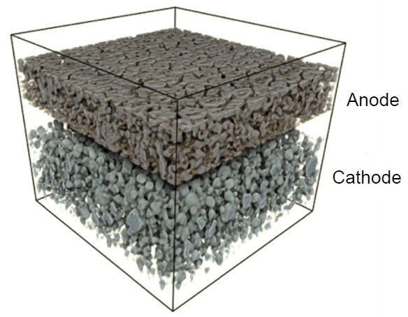
Figure 1: X-ray CT 3D rendering image 1.08 μm/voxel
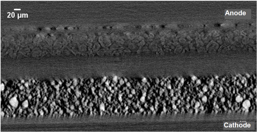
Figure 2: X-ray CT Tomography image 1.08 μm/voxel voxel
Samples provided by: Consortium for Lithium Ion Battery Technology and Evaluation Center (LIBTEC)

Contact Us
Whether you're interested in getting a quote, want a demo, need technical support, or simply have a question, we're here to help.