Submicron CT for Plant Research Applications
Introduction
X-ray submicron computed tomography (submicron CT) is a powerful method for 3D visualization and inspection of many types of samples. This non-destructive method provides sufficient resolution and contrast to evaluate any microstructural features, with the ability to resolve structures even below one micron. Moreover, this method requires minimal/no sample preparation, eliminating complicated tasks such as embedding, coating or thin slicing required with other high-resolution methods. The Rigaku nano3DX represents state-of-the-art laboratory-based nanoscale X-ray imaging. This device, when used with deep learning methods, is an unmatched tool for the plant science.
Instrument
The nano3DX is a true X-ray microscope (XRM) with the ability to measure relatively large samples at very high resolution. This is accomplished by using a high-powered rotating anode X-ray source and high-resolution sCMOS X-ray camera. The rotating anode provides for fast data acquisition and the ability to switch anode materials easily to optimize the data acquisition.
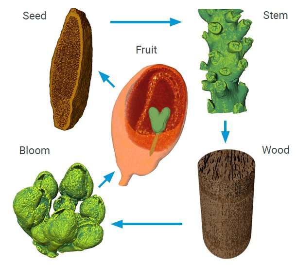
Figure 1: nano3DX allows for control over the entire life cycle of a plant.
CT data and results
With the introduction of high-resolution CT systems such as Rigaku nano3DX, plant tissue samples can be now imaged at cellular resolution in a laboratory environment.
Applications include the study of plant development and organ morphogenesis, as well as mathematical modelling of various processes such as the transport phenomena. The simplicity of sample preparation, moreover, allows for broader accessibility but also for preserving the native structures of plant specimens, therefore avoiding complicated interpretation of the results. Using Rigaku’s nano3DX, the complete information about any plant sample, from macroscale to nanoscale, can be easily acquired in a non-destructive manner, thus enabling visualization and quantification of cellular features and intracellular spaces on a daily basis.
nano3DX provides insight into key factors of plant life cycle, such as:
- Phenotype evaluation
- Structural morphology
- Tissues and cellular-features analysis
- Root systems analysis
- Seeds, fruits and embryos analysis
- Vascularization analysis
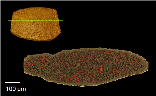
Figure 2: Morphological analysis of seed and storage substances (visualized by red color)—length of the seed along the major axis: 864 μm; width of the seed along the minor axis: 267 μm; total volume of the seed: 0.037 mm³; volume of the storage substances: 0.0019 mm³; the ratio between storage substances and the seed volume: 5.14%. Courtesy of Dr. Ulla Nauman (MPI for plant breeding research).
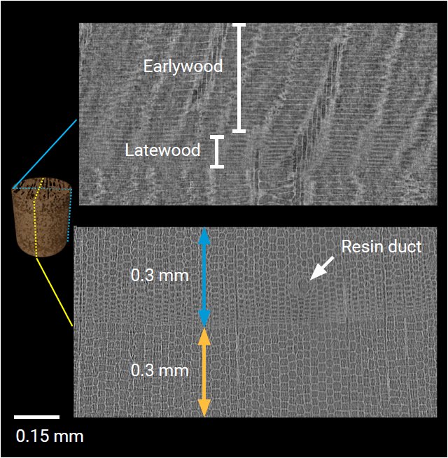
Figure 3: Wood analysis—individual growth cells forming wood rings are visible in the nano scale scan. Approximate width of one ring is 0.8 mm. According to the quantitative analysis (lower image), there are 19 cells (blue arrow) in the selected area of one ring, and 12 cells (yellow arrow) in the same area of different ring. A resin duct, marked by a white arrow is also visible. Wood cells can be divided into latewood and earlywood cells. The difference is in their density. The lateral view on the earlywood and latewood cells is depicted in the upper image. Courtesy of Jan Van den Bulcke (UGhent Woodlab, UGent Center for X-ray Tomography).
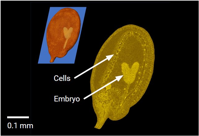
Figure 4: Fruits analysis—in the pseudo-colored cross section, individual cells forming the fruit and an embryo in the middle of the fruit are clearly visible. Courtesy of Dr. Ulla Nauman (MPI for plant breeding research).
CT analysis performed by CEITEC – CT Lab

Contact Us
Whether you're interested in getting a quote, want a demo, need technical support, or simply have a question, we're here to help.