Visualization and Size Analysis of Foreign Matter in CFRP using a High-resolution 3D X-ray Microscope
Introduction
The number and volume of voids and foreign matter in molding materials affects the strength of the material. High-resolution 3D X-ray microscopes enable 3D visualization and quantification of substances with different densities in an object 1 mm or less in size. In this example, we performed a CT scan on a carbon fiber reinforced plastic (CFRP) and analyzed the location and size distribution of foreign matter.
Measurements and results
The CT scan of CFRP was performed with a Cu X-ray source for 30 minutes. The tomographic images of the CT reconstruction results are shown in Figure 1. The images show carbon fibers, resin, voids, and foreign matter rendered with different gray scales. The foreign matter image was displayed as a 3D image (Figure 2) and the histogram of the particle size (the diameter of spheres with the same volume) of the foreign matter is shown in Figure 3. As seen from this example, high-resolution 3D X-ray microscopes allow the extraction and quantification of microscopic substances with different densities.
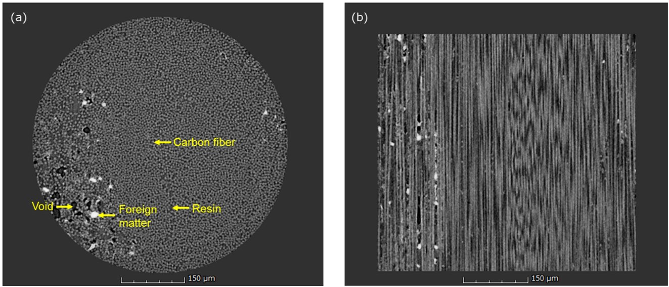
Figure 1: CT slice images of CFRP (a) Top view of the sample (b) Side view of the sample
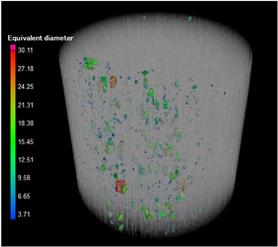
Figure 2: A CT volume rendering image of CFRP (foreign matter colored)
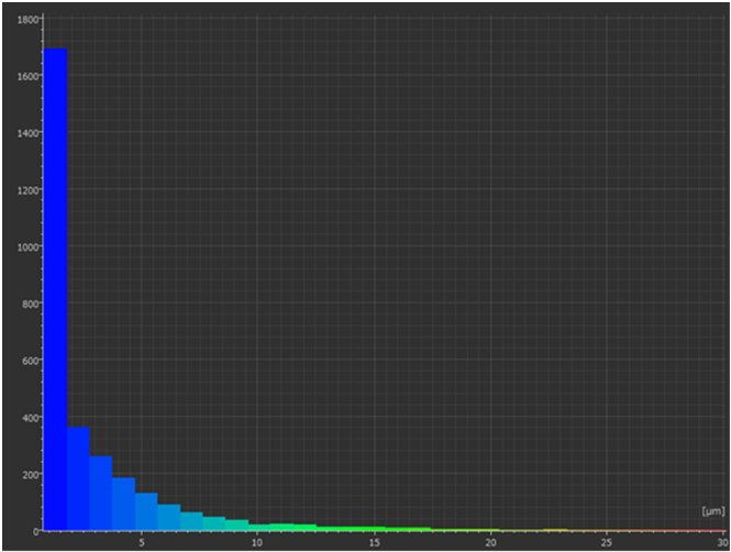
Figure 3: Particle size distribution of foreign matter

Contact Us
Whether you're interested in getting a quote, want a demo, need technical support, or simply have a question, we're here to help.