生命科學研究
從蛋白質晶體結構解析到分子成像
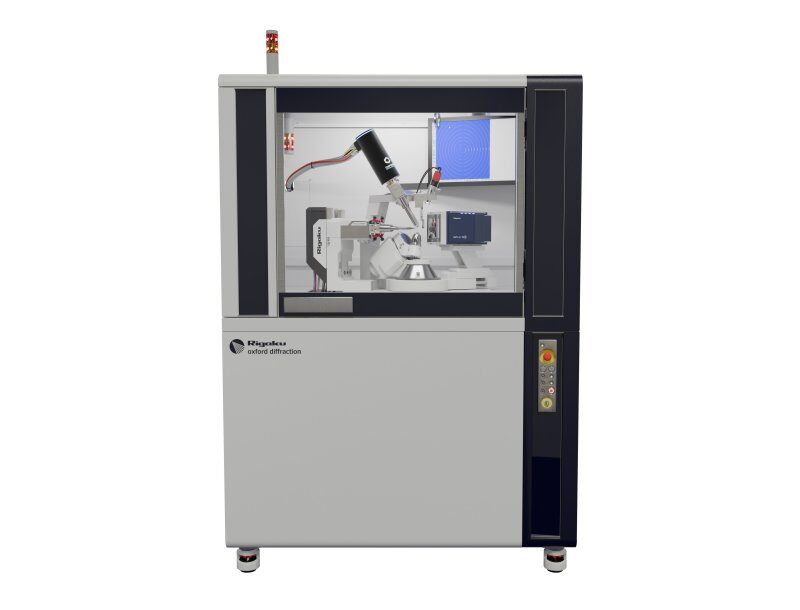
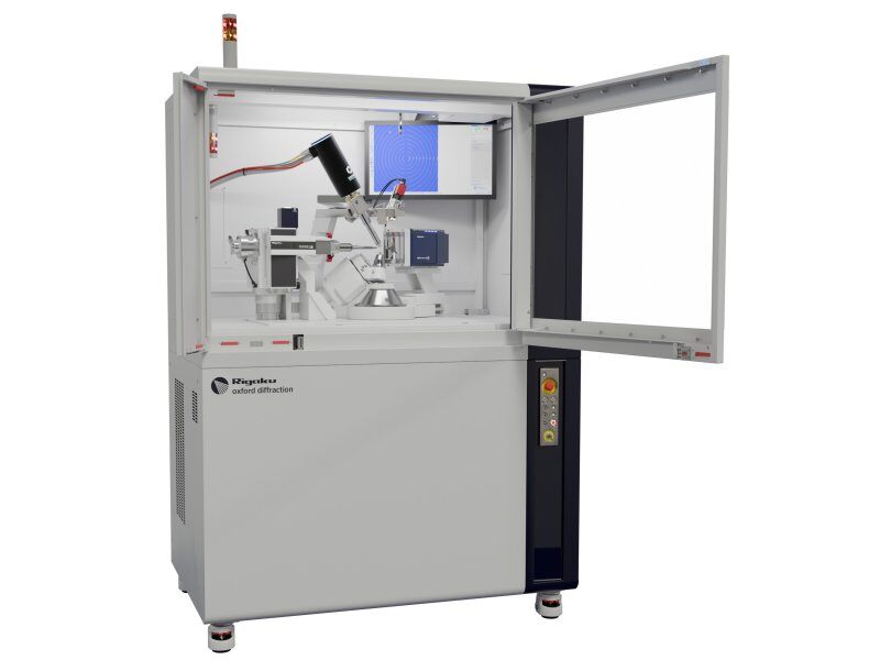
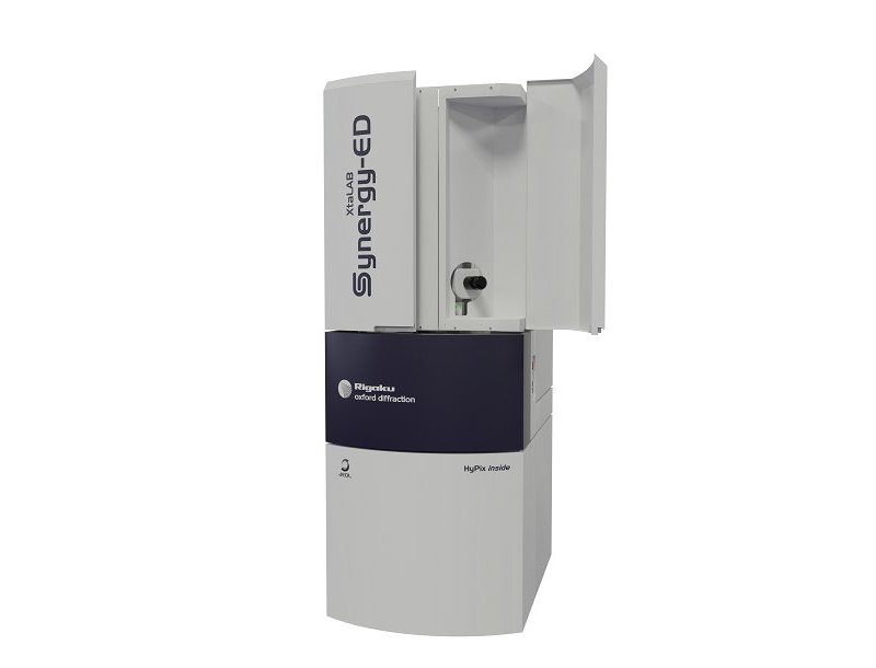
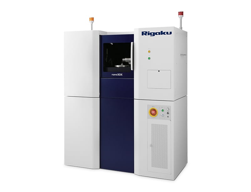
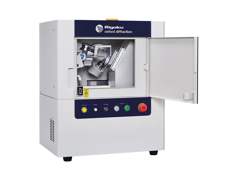
X光與電子繞射技術是解析蛋白質及小分子三維結構的關鍵工具,為藥物發現與生命科學研究提供核心支援。在臨床前研究階段,多模態分子成像系統可實現小動物器官結構與功能的精準可視化。此外,X光電腦斷層掃描(CT)技術還能夠為植物、昆蟲及軟組織等生物樣品提供高解析度無損結構表徵。
應用說明
探索成功應用案例,選出最佳分析技術
-
XCT2201 - Submicron CT for the Life Sciences: Animal and Tissue Research Applications
-
XCT2103 - Submicron CT for Plant Research Applications
-
RACCT9046 - Wild Bird Forensic
-
RACCT9024 - Tomato Plant Root System Analysis by X-ray CT
-
RACCT9023 - Maize Kernel Analysis by X-ray CT
-
RACCT9022 - Coral Surface Area and Porosity Analysis by X-ray CT
-
RACCT9021 - Cicada Analysis by X-ray CT
-
PX031 - Method: MicroED/3D ED on Proteins Using the XtaLAB Synergy-ED
-
PX030 - Using CrysAlisᴾʳᵒ to Tackle Macromolecular Non-merohedral Twins
-
PX029 - Intoducing the XtaLAB Synergy Flow
-
PX028 - Resolving Large Unit Cells by using the Correct Detector, Distance, and Beam Divergence
-
PX027 - Synergy Of PX and SMX on One Diffractometer with the HyPix-Arc 150°
-
PX026 - Sulfur SAD Phasing of Thaumatin in 10 Minutes with the HyPix-Arc 150° and XtaLAB Synergy-DW
-
PX024 - Lysozyme S-SAD Phasing with Two Minutes of Data Collection on the XtaLAB SynergyCustom
-
PX023 - ClpS SAD phasing with 30 Minutes of Data Collection on the XtaLAB Synergy-S
-
PX022 - Thaumatin S-SAD Phasing with 2 hours of Data Collection on the XtaLAB Synergy-S
-
PX021 - I-SAD Phasing of the HIV Integrase Core Domain using Data Collected on the XtaLAB Synergy-S
-
PX020 - In situ Data Collection and Structure Solution using the XtalCheck-S System
-
PX019 - Using the EIGER 4M for Single Crystal X-ray Diffraction on Catalase
-
PX018 - CrysAlisᴾʳᵒ: An All-in-one Software Package for Single Crystal X-ray Diffraction
-
PX015 - Data Collection and Improved Spot Resolution with the HyPix-6000HE HPC Detector
-
B-XRI1017 - Analysis of volume fraction and thickness of bran layer using a high-resolution 3D X-ray microscope
推薦產品
未找到心儀產品?立即體驗“產品檢索器”
MILabs - 理學旗下成員企業 分子成像
MILabs 是分子成像領域的全球引領者,致力於提供高性能臨床前多模態成像系統,支援micro-PET、micro-SPECT、micro-CT 和光學斷層掃描系統的無縫整合。