At the beginning of June we celebrated the installation of the first XtaLAB Synergy-ED at a customer site in the Americas. The finale was a ribbon-cutting ceremony with Northwestern University, Rigaku and JEOL dignitaries on hand for the occasion.
View in browser
Rigaku_Logo_RGB-1
CT Banner_05.2021

Volume 16, No. 6, June 2024

At the beginning of June we celebrated the installation of the first XtaLAB Synergy-ED at a customer site in the Americas. The finale was a ribbon-cutting ceremony with Northwestern University, Rigaku and JEOL dignitaries on hand for the occasion. Christos Malliakas, below in the center, cuts the ribbon with samurai scissors.

ribbon-cutting ceremony

We are very excited as we approach the ACA Annual Meeting in Denver. We have planned a series of activities for this year, promising fun and learning for all.


Rigaku will run online demonstrations of the XtaLAB Synergy-ED throughout the ACA conference, starting with the opening ceremony on Sunday evening. Come by our booth, #305, any time during the conference and experience crystal structures being solved on submicron grains in minutes using a dedicated electron diffractometer and CrysAlisPro.

Aca Synergy-Ed Demo Promo_compressed

From the opening reception, until we run out, we will be giving away vintage Rigaku fun-run T-shirts at our booth, #305. I am told we have only 114 T-shirts, so make sure to come early to get your favorite. Please plan to wear it to the Rigaku Mixer. For those of you who already have a Rigau fun-run T-shirt, please bring it with you and wear it at our mixer as well.


Speaking of our mixer, it will be held at The Deck at the Marriott on July 9. Of course, all will be welcome with or without a T-shirt, as long you have a ticket! And, of course, you know how to get a ticket: come by booth #305! 


Several of us have presentations at the meeting: Simon Bates, Lee Daniels, Pierre LeMaguerès, Mathias Meyer, and yours truly. Please check the book of abstracts for locations and times.


With the ACA coming up, I thought it would be nice to reintroduce/introduce the application team here at Rigaku Americas. You’ll find short bios on Pierre, Mark, Lee, and our newest member Jessica below. Emilia Götz provides the Tip of the Month, merging data from the XtaLAB Synergy-ED. 


Jeanette moved to London a couple of weeks ago to join her husband and is taking a short hiatus from reviewing books. You’ll have to suffer with me for this month’s review. 


Joe Ferrara

Product of the Month


XtaLAB mini II Benchtop Single Crystal X-ray Diffractometer


Single crystal X-ray diffraction on your benchtop 

The perfect addition to any synthetic chemistry laboratory, the XtaLAB mini II single crystal X-ray diffractometer will enhance research productivity by offering affordable structure analysis capability without the necessity of relying on a departmental facility. With the XtaLAB mini II benchtop diffractometer, you no longer have to wait in line to determine your structures. Instead, your research group can rapidly analyze new compounds as they are synthesized in the lab without having to queue up in the departmental core facility. 

Rigaku TOPIQ Webinars

Rigaku has developed a series of 20–30 minute webinars that cover a broad range of topics in the fields of X-ray and electron diffraction, X-ray fluorescence and X-ray imaging. You can watch recordings of our past sessions here.

Upcoming Webinar:

TOPIQ | The Support of Structural Science at Yale University

Thursday, July 25, 2024 at 9 AM CDT / 10 AM EDT


In this webinar, Brandon Q. Mercado, Ph.D. will explore the comprehensive support for the advancement of structural science provided by The Chemical and Biophysical Instrumentation Center in the Chemistry Department at Yale University. His facility is equipped with state-of-the-art instrumentation, including single crystal X-ray diffractometers, powder diffractometers, small/wide-angle X-ray scattering systems, and micro-computed tomography (microCT). These resources enable scientists to conduct detailed analyses of crystalline structures, powders, and complex materials, facilitating a broad range of scientific investigations. Additionally, he is excited to announce the upcoming addition of an electron diffractometer, which will further enhance our capabilities. Brandon will share preliminary results obtained in collaboration with Rigaku and discuss the relevance to ongoing research projects, demonstrating the potential to obtain high-resolution, impactful structural data with electron diffractometers. This presentation aims to highlight the pivotal role his core facility plays in supporting cutting-edge structural science, fostering innovation, and driving scientific discovery within the university setting.

Register now >


Upcoming Events:


American Crystallographic Association Annual Meeting, July 7-12, Denver, CO


38th Annual Symposium of The Protein Society, July 23-26, Vancouver, BC.


Denver X-ray Conference, August 5-9, Denver, CO


European Crystallographic Meeting 34, August 26-31, Padova, Italy


Second meeting of the Latin American Crystallographic Association (LACA), October 23-27, Mérida, Mexico

 
Follow Us on X (Twitter)

To keep up to date on the latest news and events from Rigaku Oxford Diffraction, follow our Twitter feed.

Follow


Join Us on LinkedIn

Our LinkedIn group 

shares information and fosters discussion about X-ray crystallography and SAXS topics. Connect with other research groups and receive updates on how they use these techniques in their own laboratories. You can also catch up on the latest newsletter or Rigaku Journal issue. We also hope that you will share information about your own research and laboratory groups.

Join Here


Rigaku X-ray Forum

At rigakuxrayforum.com you can find discussions about software, general crystallography issues and more. It’s also the place to download the latest version of Rigaku Oxford Diffraction’s CrysAlisPro software for single crystal data processing.

Join Here
XtaLAB mini II 

XtaLAB mini II Overview


Teach single crystal X-ray diffraction through hands-on experience

In many universities, the departmental single crystal X-ray diffractometer is considered off limits to students because of fear the instrument might be damaged by inexperienced users. The XtaLAB mini II benchtop diffractometer provides the opportunity for students to learn single crystal X-ray analysis by actually using a fully functional diffractometer. This is not a black box instrument. Rather, performing the important steps of mounting a crystal on the goniometer and physically centering the crystal in the position of the X-ray beam ensures that students learn the importance of mounting techniques and crystal selection. The simple design of the XtaLAB mini II X-ray diffractometer minimizes the possibility of students misaligning or damaging the system. 


Compact size with excellent X-ray data quality 

The Rigaku XtaLAB mini II is a research-grade chemical crystallography diffractometer that sits on the benchtop. No data quality compromises. Results delivered are unambiguous. X-ray source tube lifetime is extended by running a standard off-the-shelf X-ray tube at 600 W. To compensate for running at a lower power, a SHINE optic (special curved monochromator) is utilized to produce usable X-ray flux comparable to a standard X-ray diffractometer. In addition, the XtaLAB mini II is equipped with a state-of-the-art two-dimensional semiconductor detector that is characterized by extremely low background noise and high sensitivity. These detector characteristics mean that, for weakly diffracting samples, longer scan rates can be employed without the weak diffraction signals being swamped by detector noise. 


Dedicated to producing publication quality single crystal X-ray structures 

The chief design requirement when creating the XtaLAB mini II single crystal X-ray diffractometer was that the structures produced would be publishable in the most demanding scientific journals. The HPC X-ray detector is positioned so that the maximum 2θ value is well outside of the Acta Cryst. requirements. The software provides all the tools you need to generate publication quality data that can be used to determine 3D structures from a variety of structure analysis packages. 


CrysAlisᴾʳᵒ single crystal X-ray diffraction software 

The XtaLAB mini II comes complete with CrysAlisPro, our user-inspired data collection and data processing software for single crystal analysis. Designed around an easy-to-use graphical user interface, CrysAlisPro can be operated under fully automatic, semi-automatic or manual control. CrysAlisPro combines automated crystal screening, the fastest and most accurate strategy software available, concurrent data reduction and automatic small molecule structure solution. Visual feedback is provided for each step with clear, color-coded guidance so that both novices and experts can collect high-quality data in the shortest time possible. 


AutoChem: Automated single crystal structure determination software (optional) 

AutoChem is an optional software package for the XtaLAB mini II and is the ultimate productivity tool for chemical crystallographers, offering fast, fully automatic X-ray structure solutions and refinement during data collection. Developed in collaboration with OlexSys Ltd (Durham University, UK), AutoChem works in conjunction with Olex² where more advanced structure solution and refinement functionality exists. AutoChem is seamlessly integrated within CrysAlisPro and forms an integral part of our ‘What is this?’ feature. The ‘What is this?’ feature gives you structures quickly and ensures you are not wasting time collecting full datasets on known samples or starting materials. It is an alternative pre-experiment option, which is used to plan your full data collection. 

Application Lab in the Spotlight

Pierre LeMaguerès

Pierre LeMaguerès is Director of Scientific Support at Rigaku Americas. Having spent many years as an application scientist, in both protein and small molecule X-ray crystallography, Pierre now manages the single crystal department at Rigaku. The group of crystallographers Pierre manages uses state-of-the-art Rigaku X-ray diffractometers to support customers, engage in the development of new hardware and software products for single crystal crystallography and help the Sales and Business Development departments develop relationships with new partners in key markets. 

Mark Del Campo

Mark Del Campo is a Senior Applications Scientist at Rigaku Americas with over 20 years of experience in the life sciences. He supports Rigaku’s macromolecular crystallography, small angle X-ray scattering, and microED customers. He has traveled the world supporting Rigaku’s customers for 13 years. Mark did his postdoctoral research on fungal DEAD-box proteins and Group I and Group II introns with Dr. Alan Lambowitz at the University of Texas at Austin, where he solved eight structures deposited in the PDB. Mark received his Ph.D. under the supervision of Dr. James Ofengand at the University of Miami, where he worked on E.coli pseudouridine synthases and solved three structures deposited in the PDB. Mark was the recipient of both predoctoral and postdoctoral Ruth L. Kirschstein National Research Service Awards. 

Lee Daniels

Lee Daniels is a senior applications scientist for Rigaku Americas. Lee spent 15 years as an academic crystallographer and a Rigaku user before joining Rigaku for the first time over 20 years ago. Lee was first exposed to crystallography while working towards his PhD under Prof. Frank Albert Cotton at Texas A&M University. Following two years in Zeigler-Natta catalysis research at Dow Chemical, he returned to a crystallography post-doc before starting the service crystallography lab at Iowa State University. He later returned to Texas A&M to manage the crystallography and computing laboratories in F.A. Cotton's research facilities while teaching crystallography and training scores of students and post-docs. He then joined Rigaku in 2002, and more recently spent some time representing the Cambridge Crystallographic Data Centre in North America before returning to his role at Rigaku. 

Jessica Burch

Jessica Burch recently joined our team as an application scientist. She obtained her Ph.D. from UCLA, where she specialized in organic synthesis and the use of electron diffraction to determine the structures of small molecules. Prior to joining us, she was a staff scientist at Caltech, focusing on methods development in electron diffraction. 

Crystals in the News

May 22, 2024

Scientists from ORNL and NIST have synthesized and characterized a 147Pm+3 compound in solution and the solid state, helping to complete our understanding of lanthanide chemistry.


May 22, 2024

Researchers from the UK have synthesized and characterized a series of porous isoreticular non-metal organic frameworks that can absorb molecular guests in quantities larger than conventional MOFS.


May 23, 2024

Researchers from the US (95% from Baylor College of Medicine in Houston) have developed a reversible male contraceptive by targeted inhibition of serine/threonine kinase 33.

TIP OF THE MONTH


Profit Merge 


Why merge?

Merging datasets can be helpful to enhance the quality of datasets, particularly by addressing issues of incompleteness. This is especially important for crystals measured at high pressure in diamond anvil cells or those that lose crystallinity due to beam sensitivity during measurement. In three-dimensional electron diffraction (3DED), merging becomes crucial for crystals with low symmetry, which often results in low completeness because tilting is limited to one axis only. Beyond improving completeness, merging datasets can also increase redundancy and improve atomic displacement parameters, especially for 3DED datasets. 


How to merge in CrysAlisPro

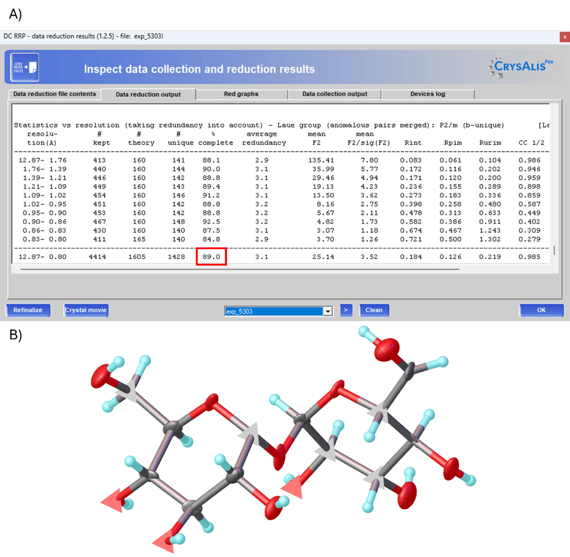
If the measurement of a crystallite yields insufficient data for a satisfactory structure solution (Fig. 1A), the dataset can be merged. In this example, the dataset was collected via 3DED with an XtaLAB Synergy-ED. Due to the possibility of tilting along one axis only, the completeness of the monoclinic structure is limited. The initial dataset has a completeness of 89%. While the structure could be solved using Olex2, the refined structure displays numerous negative ADPs (Fig. 1B).

CrysAlisPro Proffit Merge 1

Figure 1: A) Data reduction results of a 3DED dataset with limited completeness due to its low symmetry. B) Refined structure of the dataset shown in A) with numerous negative ADPs. 


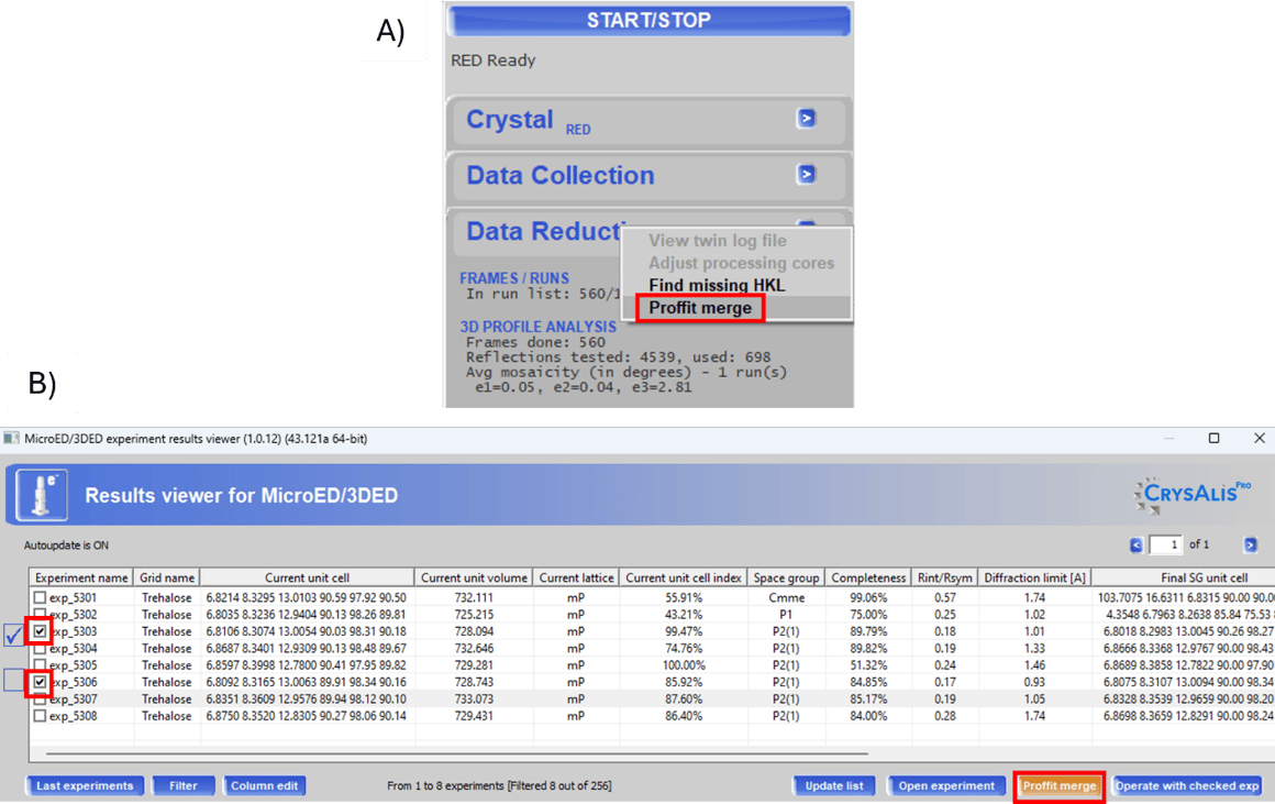
In CrysAlisPro, the function to merge datasets can be accessed in several ways. One method is to click on the arrow in the Data Reduction tab on the right side of the user interface and select [Proffit merge] (Fig. 2A). For 3DED datasets, this can also be done through the Results Viewer by selecting the desired datasets and clicking on [Proffit merge] (Fig. 2B).

CrysAlisPro Proffit Merge 2

Figure 2: Different ways of accessing Proffit merge. The window can be opened either via the Data Reduction tab (A) or the Results Viewer (3DED only) (B). 


For both options, the Proffitmerge dialog will open where .rrpprof files of the desired datasets can be loaded using the [Add] button. Once a dataset is added, detailed information such as the unit cell, crystal system and volume will be displayed. The bottom half of the window shows the overall completeness and minimum d-spacing (dmin) (Fig. 3A). You can add as many datasets as desired, provided they are in the same space group and have the same arrangement of cell axes. The unit cells should be as similar as possible (See Fig 3B). Additionally, the datasets should have comparable intensity ranges. Merging datasets with very strong and very weak data can introduce large errors in the weaker data due to the scaling during the merging process. Datasets can be removed using the [Delete] button. Once the completeness is satisfactory, you can choose to [Create a script] or [Create and run script] to save a .mac file or to save and execute it, respectively. Here, a name and directory can be specified. Alternatively, by clicking [OK], the .mac file is automatically saved and run in the current directory (if box is checked) with the name specified in the Output file name window. By default, this name is a combination of the merged experiments.

CrysAlisPro Proffit Merge 3

Figure 3: Proffitmerge dialog. Experiments can be added and removed from the list of datasets to be merged. The overall completeness is shown. 


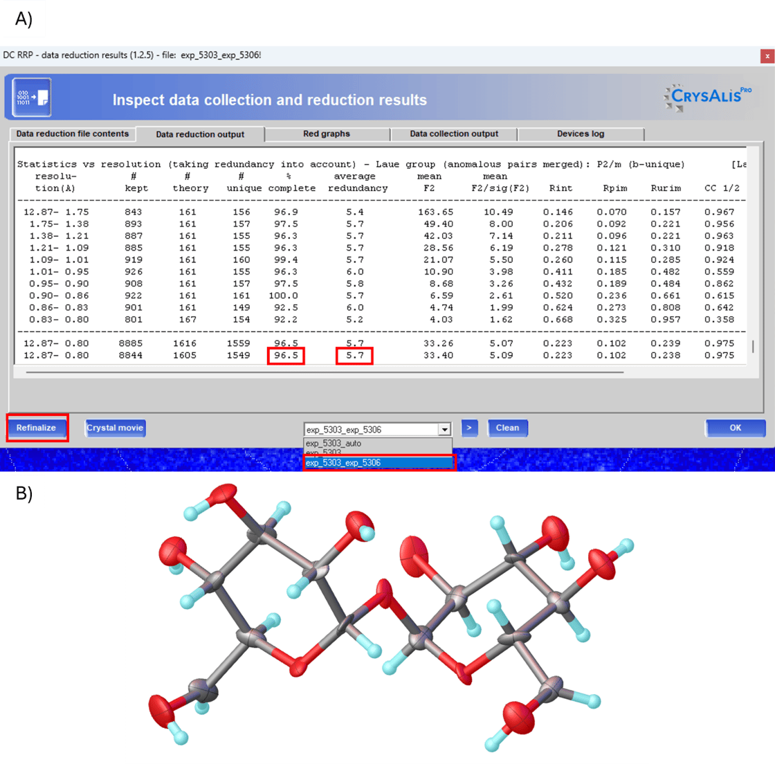
Afterwards, select the newly generated merged dataset in the Data reduction results and refinalize it. In this example, compared to the single dataset, the completeness has increased from 89% to 96.5% and the redundancy has improved from 3.1 to 5.7 (Fig. 4A). Additionally, the ADPs in the refined structure are now all positive definite (Fig. 4B).

CrysAlisPro Proffit Merge 4

Figure 4: A) Data reduction results of the merged 3DED datasets with higher completeness and redundancy. B) Refined structure of the merged dataset shown in A) with improved ADPs. 

Book Review

Review: The Scientific Truth, the Whole Truth and Nothing but the Truth 

By John R. Helliwell

ISBN 9781032521398


book cover

John R. Helliwell is well known to this community for his contributions to the field of crystallography, as well as being a dedicated educator and mentor, contributing to the training and development of the next generation of scientists. His commitment to the scientific community is reflected in his extensive publication record and his active participation in various scientific organizations and conferences.


His book emphasizes the distinction between accuracy and precision in scientific measurements. While precision refers to the consistency of repeated measurements, accuracy relates to how close a measurement is to the true value. My friend and former colleague Jim Pflugrath would also add that accuracy trumps precision.


John also discusses the importance of pragmatic approaches in scientific research. Sometimes, practical considerations (such as available resources or time constraints) influence the choice of methods and measurements. In keeping with the significant changes in structural science brought by AI, the book explores the application of machine learning and artificial intelligence to crystallography databases in biology and chemistry. These technologies can enhance data analysis and accelerate scientific discoveries. 


This book is written for scientists at all stages of their careers but is directed at younger scientists. My only negative comment is that the book is rather expensive and will put a dent in a student’s limited budget. Even so, this book is particularly relevant for early career scientists and university students. It provides insights into scientific methodology, critical thinking, and the pursuit of truth. 


Review by Joe Ferrara


Subscribe to Rigaku newsletters!

X
LinkedIn
Facebook
Instagram
YouTube

© 2024 — Rigaku Holdings Corporation and its Global Subsidiaries. All rights reserved.

Rigaku Americas - A Division of Rigaku Americas Holding, Inc.s, Inc. 9009 New Trails Dr.The Woodlands, TX 77381 United States +1 281 362-2300


Manage subscription preferences or unsubscribe