Backgrouond
With the MiniFlex, it is possible to install a graphite monochromator for a scintillation counter, of the type used in high-grade, standalone, general-purpose equipment. By using a monochromator, it is possible to obtain an X-ray diffraction pattern with high P/B ratio because the monochromator can remove interfering components such as continuous X-rays produced by the X-ray tube, Kβ-rays, and fluorescent X-rays from the sample.
Investigation
Fig. 1 shows the X-ray diffraction pattern for hematite, measured using the Kβ filter method and the graphite monochromator method. The large reduction in background makes it possible to observe very weak peaks as illustrated by the inset box between 38 and 40°. This makes identification of trace phases much easier.
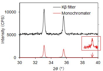
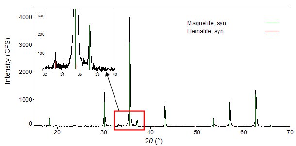
Fig. 2 shows the X-ray diffraction pattern and qualitative analysis results for Fe oxide, measured using a graphite monochromator. By using a graphite monochromator, it is possible to obtain an X-ray diffraction pattern with an extremely low background, and thereby detect trace components.
Apparatus condition:
MiniFlex600 (F.F tube 40 kV 15 mA), Detector: Scintillation counter (monochromater), Slit conditions: DS / SS = 1.25°, RS = 0.3 mm, Incident side and receiving side Soller slit: 5°, Incident height limiting slit = 10 mm
Measurement condition:
Scan range: 2θ = 10 ~ 70°, Step width: 0.02°, Scan speed: 4° / min. (about 15 min.)